

A lot of students hit the same wall in nephrology. You open a board-style vignette, see creatinine, urine sodium, edema, pH, potassium, and a vague history of diabetes or vomiting, and suddenly it feels like five subjects got stacked into one question.
The fastest way to make nephrology manageable is to stop treating it like isolated facts. For exams, nephrology becomes much easier when you build it in layers: renal physiology first, then urinalysis patterns, then electrolytes and acid-base reasoning, then disease recognition. That's the approach here.
Byline: Ace Med Boards Editorial Team
Why Nephrology Feels Hard and How This Guide Helps
You are 45 seconds into a board-style vignette. A patient has rising creatinine, leg edema, a low bicarbonate, and a medication list that includes an ACE inhibitor and ibuprofen. If you try to solve each clue separately, the question feels crowded fast. If you read it as one kidney story, the stem becomes much easier to organize.
Nephrology feels hard because the kidney is less visible than a failing pump or an obstructed airway. You cannot watch filtration happen. You have to infer it from labs, urine findings, volume status, and timing. For a second-year student, that often feels like being asked to diagnose a plumbing problem by listening through the wall.
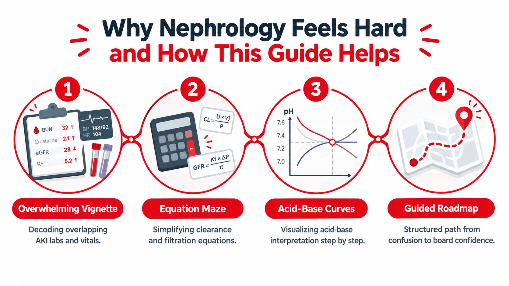
A more useful way to frame the subject is this: nephrology is physiology under pressure. Board questions keep asking the same sequence. What is the kidney sensing. Which response should follow. Where in the nephron is that response happening. Which disease pattern or drug effect could disrupt it.
That sequence is the backbone of this guide.
Why students get stuck
The struggle usually comes from three predictable habits.
- Memorizing categories without links: It is easy to learn nephritic syndromes, nephrotic syndromes, diuretics, and acid-base rules as separate charts. The exam then blends them into one vignette.
- Learning formulas before mechanisms: A compensation rule sticks better when you know what the lungs and kidneys are trying to correct.
- Reading single values instead of trends: Creatinine, urine output, potassium, and blood pressure make more sense when followed across time.
A simple checkpoint helps: ask what the kidney is trying to keep, what it is trying to dump, and what signal pushed it in that direction.
How this guide makes the subject easier
This article is built from physiology outward, then tied back to exam reasoning. Each major topic is paired with a board-style clinical vignette, and composite examples are labeled clearly so they teach pattern recognition without pretending to be real student outcomes.
That matters because nephrology is one of the easiest subjects to study passively and one of the hardest to answer under time pressure. Short rounds of active recall practice for medical studying work especially well here because they force you to retrieve nephron functions, diuretic targets, and acid-base steps in the same way the exam will.
The payoff is larger than test performance alone. Kidney disease touches a huge patient population worldwide, and the clinical burden continues to grow, as noted earlier in the article. So the goal here is not to drown you in renal trivia. It is to help you build a repeatable method: start with physiology, trace the disturbance, and match it to the vignette in front of you.
Foundations of Renal Physiology for the Boards
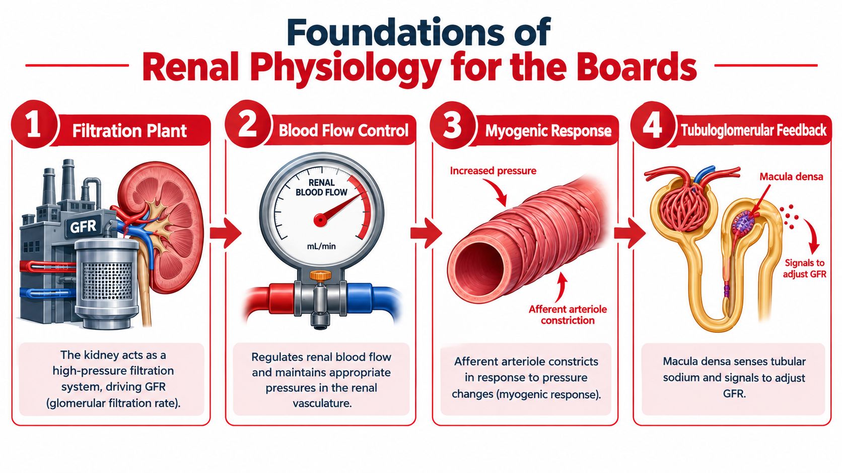
Think of the kidney as a high-pressure filtration plant attached to a very selective recycling system. The glomerulus filters. The tubules decide what the body keeps and what it throws away.
That image is much more useful than trying to memorize every transporter on day one.
Start with glomerular filtration rate
Glomerular filtration rate (GFR) is the amount of plasma filtered across the glomerulus per unit time. On exams, the key question isn't “What is the formula?” nearly as often as “What would raise or lower filtration pressure?”
A helpful mental model:
- Afferent arteriole: controls blood entering the filter
- Efferent arteriole: controls blood leaving the filter
- Glomerulus: the filter membrane itself
If the afferent arteriole constricts, less blood enters, so GFR tends to fall. If the efferent arteriole constricts mildly, pressure can back up into the glomerulus and support filtration. That's why drugs that alter arteriolar tone matter so much in board questions.
Autoregulation makes the kidney less fragile than it looks
The kidney doesn't passively accept every blood pressure swing. It uses myogenic response and tubuloglomerular feedback.
The easiest analogy is a sink with an automatic valve. If pressure rises too much, the incoming pipe tightens. If sodium delivery to the macula densa changes, the nephron sends a signal to adjust upstream flow.
When a stem mentions nonsteroidal anti-inflammatory drugs, angiotensin-converting enzyme inhibitors, dehydration, or renal artery stenosis, test writers are usually asking whether autoregulation can still protect GFR.
Know the nephron segment jobs
Most board questions become easier if you can say the main job of each segment in one line:
- Proximal tubule: bulk reabsorption. Sodium, water, glucose, amino acids, bicarbonate.
- Loop of Henle: creates the medullary gradient. The thick ascending limb reabsorbs solute and is water-impermeable.
- Distal convoluted tubule: fine-tunes electrolytes.
- Collecting duct: final water and sodium decisions under hormonal control.
Antidiuretic hormone (ADH) helps the collecting duct reabsorb water. Aldosterone promotes sodium reabsorption and potassium secretion in the distal nephron. Atrial natriuretic peptide (ANP) pushes the system in the opposite direction by favoring natriuresis.
A loop diuretic question is really a question about the thick ascending limb. An angiotensin-converting enzyme inhibitor question is often really a question about efferent arteriolar dilation and filtration pressure. If you want a focused content review after this overview, the Ace renal physiology page is one option for organizing those mechanisms alongside question practice.
AKI and CKD From Definition to Diagnosis
A board stem gives you a patient with a creatinine of 2.1 mg/dL. The number looks alarming, but by itself it answers very little. Your first job is to place that value on a timeline. Did kidney function fall over hours to days, or has abnormal function been present for months?
That is the starting split between acute kidney injury (AKI) and chronic kidney disease (CKD).
CKD means kidney damage or reduced kidney function present for more than 3 months. For classification, use the CGA framework: Cause, GFR category, and Albuminuria category. The GFR categories are G1 (≥90), G2 (60 to 89), G3a (45 to 59), G3b (30 to 44), G4 (15 to 29), and G5 (<15 mL/min/1.73 m²). Albuminuria categories are A1 (<30 mg/g), A2 (30 to 300 mg/g), and A3 (>300 mg/g) (American Family Physician CKD review).
Students often get stuck because AKI and CKD can coexist. A patient may have diabetic CKD at baseline, then develop volume depletion, sepsis, obstruction, or nephrotoxin exposure on top of it. On exams, that mixed picture is common because it tests whether you can separate background disease from the new hit.
Start with the clinical question
For AKI, ask: what changed recently that could lower GFR or injure the nephron?
For CKD, ask: what abnormal pattern has persisted long enough to count as chronic disease, and how is it classified?
That reasoning pattern is more useful than memorizing isolated creatinine cutoffs.
KDIGO's current CKD guidance also stresses following trends over time rather than reacting to one value in isolation. In a patient already known to have CKD, a meaningful shift in kidney function deserves evaluation in clinical context, and ongoing follow-up should include both GFR and albuminuria, as noted earlier in the article.
AKI Versus CKD Diagnostic Comparison
| Feature | AKI | CKD |
|---|---|---|
| Time course | Sudden change over a short period | Persistent abnormality for more than 3 months |
| Main exam question | What new process lowered kidney function? | What chronic pattern is present and how is it staged? |
| Urinalysis role | Helps localize cause quickly | Helps classify chronic damage and detect ongoing activity |
| Imaging clues | Obstruction may need urgent recognition | Small echogenic kidneys can support chronicity |
| Common board trap | Missing acute-on-chronic injury | Labeling every elevated creatinine as “renal failure” without classification |
A labeled composite vignette
Composite example: A 72-year-old with longstanding hypertension and diabetes presents with three days of vomiting and poor oral intake. Creatinine is above baseline. Urine is concentrated, and the sediment is bland.
Work through it in order.
First, this patient may already have CKD risk factors. Second, the recent vomiting creates a plausible trigger for AKI from decreased effective perfusion. Third, the urinalysis does not suggest an active inflammatory or tubular sediment. That combination makes pre-renal AKI superimposed on possible CKD the best first frame.
Now change one detail. The same patient had prolonged hypotension after sepsis, and the urinalysis shows muddy brown granular casts. Your leading diagnosis should shift toward acute tubular injury, which is an intrinsic renal cause of AKI.
This is the exam habit to build: do not ask only whether the creatinine is high. Ask what bucket the story fits.
The three AKI buckets
- Pre-renal: perfusion problem. The kidney is structurally trying to conserve sodium and water.
- Intrinsic: nephron tissue is injured. Urine sediment often gives the clue.
- Post-renal: urine outflow is blocked. History, bladder findings, or imaging may point you there.
A simple analogy helps here. AKI sorting works like tracing a plumbing problem. Pre-renal means not enough water is reaching the filter. Intrinsic means the filter or tubules are damaged. Post-renal means the drain is blocked.
If the stem also gives bicarbonate, pH, or PaCO2, keep the acid-base problem organized separately from the kidney timeline. A quick review of how to calculate and interpret the anion gap can help you avoid merging two different questions into one.
Glomerular Disease Patterns Made Clear
A board stem gives you a patient with puffy eyelids, cola-colored urine, and a recent sore throat. Another gives you leg edema, foamy urine, and a normal blood pressure. Both point to the glomerulus, but they are asking different questions. Your first job is pattern recognition.
Start with two buckets: nephritic and nephrotic.
That split works because the glomerulus has two main jobs. It filters water and small solutes, and it keeps cells and large proteins in the bloodstream. If inflammation injures the capillary wall, red cells leak across and the filtration rate often falls. That creates a nephritic picture. If the filtration barrier becomes unusually permeable to protein, albumin spills into urine and plasma oncotic pressure drops. That creates a nephrotic picture.
A useful comparison is a coffee filter. If the filter becomes inflamed and torn, you see debris and impaired flow. If its charge barrier fails, protein slips through even when the structure still looks partly intact.
Start with the urine and the story
Nephritic syndromes usually point toward glomerular inflammation. Look for hematuria, red blood cell casts, hypertension, and azotemia.
Nephrotic syndromes point toward heavy protein loss through the filtration barrier. Look for marked proteinuria, edema, and low serum albumin.
Red blood cell casts matter because they localize bleeding to the nephron rather than the lower urinary tract. Foamy urine matters because it hints at protein, not blood. If a stem gives both blood and substantial protein, keep a mixed pattern in mind rather than forcing the case into one pure category.
One exam habit helps a lot here. Read the urinalysis before you commit to the diagnosis. An active sediment with blood and casts should push glomerulonephritis high on your list.
Protein on dipstick also needs confirmation with a quantitative urine measure, as noted earlier in the article. For exam purposes, that means a positive screening test is often the beginning of the workup, not the endpoint.
High-yield patterns the boards like to test
| Disease | Pattern | Key UA Finding | Complement | Classic Association |
|---|---|---|---|---|
| IgA nephropathy | Nephritic | Hematuria, often with red blood cells | Usually normal | Hematuria after mucosal infection |
| Poststreptococcal glomerulonephritis | Nephritic | Hematuria, active sediment | Often low | Recent streptococcal infection |
| Membranoproliferative pattern | Mixed or nephritic-nephrotic | Protein and blood may both appear | Often low | Complement-mediated or immune complex patterns |
| Focal segmental glomerulosclerosis | Nephrotic | Heavy proteinuria | Usually normal | Podocyte injury pattern |
| Membranous nephropathy | Nephrotic | Heavy proteinuria | Usually normal | Adult nephrotic syndrome |
| Diabetic nephropathy | Nephrotic-range proteinuria can occur | Albuminuria | Usually normal | Longstanding diabetes |
How to reason through these patterns on an exam
Use a simple sequence.
- Identify the dominant urine pattern. Blood and casts suggest nephritic disease. Heavy protein suggests nephrotic disease.
- Check the complement clue. Low complement narrows the field. Normal complement does too.
- Match the time course and association. Infection timing, diabetes history, or edema pattern often gives the last clue.
- Ask whether the stem is hinting at a biopsy-level diagnosis or only a syndrome. Some questions want the syndrome first, not the exact histology.
Composite vignette 1: A patient develops gross hematuria within a day of an upper respiratory infection. Complement is normal. This pattern fits IgA nephropathy better than postinfectious glomerulonephritis, because IgA classically occurs close to the mucosal infection.
Composite vignette 2: A child presents with periorbital edema, hypertension, cola-colored urine, and low complement after a recent streptococcal infection. That pattern supports poststreptococcal glomerulonephritis.
Composite vignette 3: An adult has progressive edema, heavy proteinuria, and relatively bland urine sediment. That is a nephrotic syndrome pattern. The next step in board reasoning is to sort likely causes such as membranous nephropathy, FSGS, or diabetic kidney disease using the rest of the stem.
When the question is really asking about urgency
Some stems are less about naming the exact disease and more about recognizing a pattern that needs specialist evaluation. Pay attention when you see:
- Rapidly progressive kidney dysfunction
- Nephrotic-range protein loss
- Unexplained acute kidney injury with active sediment
- Suspicion for rapidly progressive glomerulonephritis
Crescents on biopsy, a fast creatinine rise, or systemic features such as pulmonary symptoms should make you think about aggressive glomerular injury, even if the stem gives limited pathology detail.
A final board-level nuance: some patients with chronic kidney disease do not fit neatly into the usual etiologic categories. As noted earlier, recent reviews describe a CKDx framework that uses genetic and histologic testing for kidney disease of unexplained cause. For exams, the practical lesson is simpler. If the pattern does not fit the common buckets, do not force it. Recognize uncertainty, then choose the next best diagnostic step.
Electrolytes and Acid-Base Step by Step
Electrolytes and acid-base questions feel intimidating because students often try to solve them all at once. Don't. Use an order.
Sodium first means water first
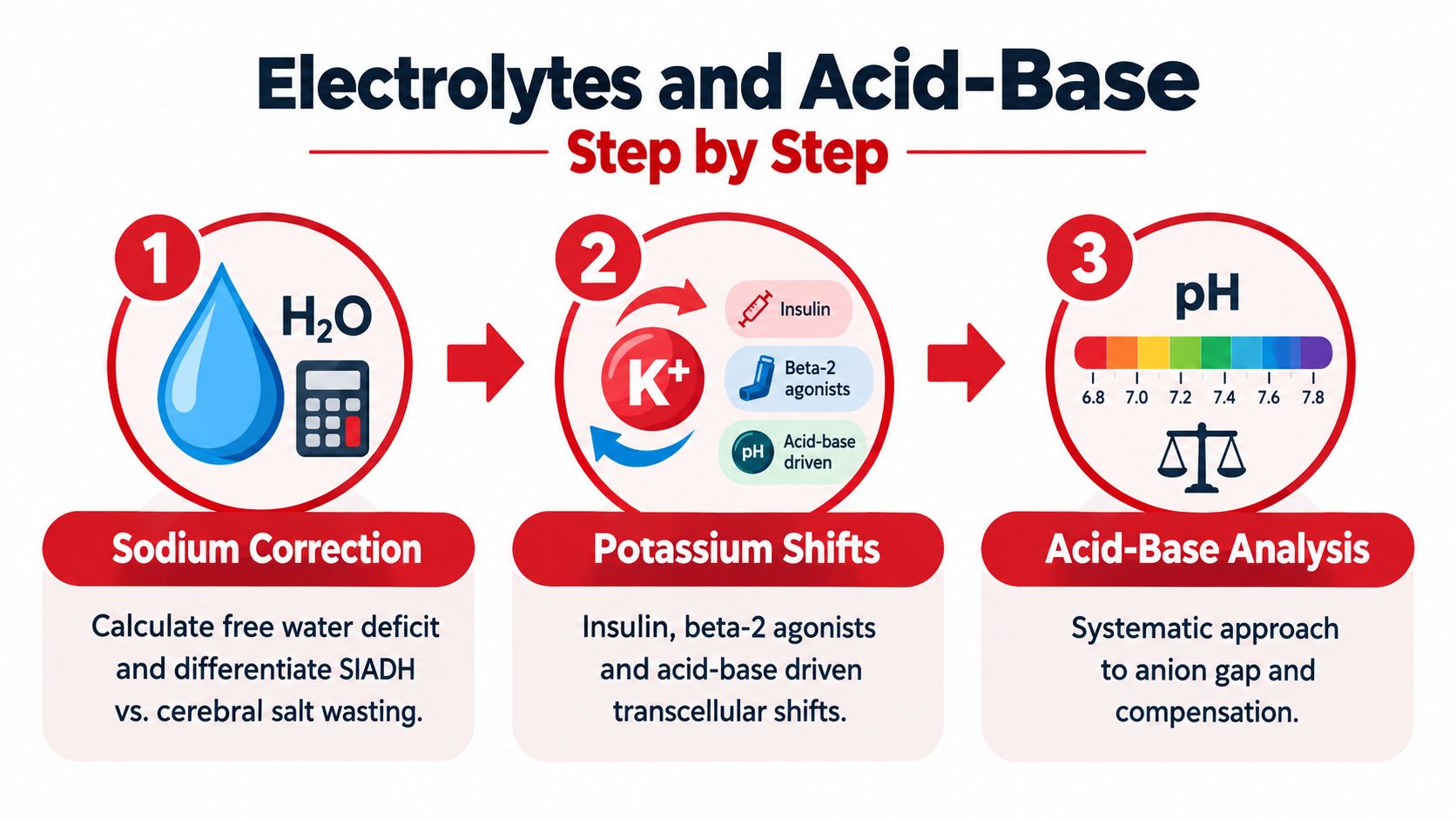
A sodium disorder is usually a water problem before it's a salt problem. If sodium is low, ask whether the patient is hypovolemic, euvolemic, or hypervolemic.
A practical board sequence:
- Check serum sodium
- Decide volume status from the vignette
- Use urine clues to see whether the kidney is appropriately conserving sodium and water
- Link the pattern to mechanism
Worked composite example: A patient with vomiting, orthostasis, dry mucous membranes, and hyponatremia most likely has hypovolemic hyponatremia. The body senses low effective circulating volume, raises antidiuretic hormone, and retains water. The low sodium is real, but the trigger is volume depletion.
Potassium questions are usually asking where potassium is
Serum potassium reflects both total body potassium and transcellular shifts. That's why a value can change quickly even before total body stores fully change.
Think in three buckets:
- Shift into cells: insulin, beta-2 agonism
- Shift out of cells: some acid-base disturbances and cell breakdown states
- Impaired excretion: reduced kidney function or altered distal nephron handling
That framing is more useful than memorizing isolated associations.
A short visual review can help before you practice questions:
Acid-base becomes manageable when the order never changes
Use the same four-step approach every time:
- Look at pH
- Identify the primary process as metabolic or respiratory
- Calculate the anion gap
- Check expected compensation, then look for a mixed disorder if things don't fit
If your acid-base answers change every time you recalculate, the problem usually isn't the formula. It's the order. Keep the order fixed.
Composite worked example: A patient with diabetes presents with tachypnea, abdominal pain, and confusion. The pH is low and bicarbonate is low, so start with metabolic acidosis. Then calculate the anion gap. If the gap is high, ask whether the respiratory compensation is appropriate. If measured carbon dioxide doesn't match expected compensation, suspect a second process.
That's the kind of reasoning pattern boards reward. A good ABG answer isn't flashy. It's disciplined. If you want more repetition on that workflow, this arterial blood gas interpretation guide is a useful companion.
One acid-base caution for real-world thinking
Board stems often simplify acid-base disorders into clean categories. Real patients frequently have mixed processes, changing volume status, and evolving kidney function. Use exam mnemonics, but don't let the mnemonic replace the physiology.
This article is for exam education and not individualized medical advice.
Exam-Specific Study Strategy and Resource Comparison
You finish a renal block question set and notice a pattern. On one exam, the stem asks you to predict how a loop diuretic changes calcium handling. On another, the same kidney topic appears as an older adult with edema, rising creatinine, and a urinalysis that points to the diagnosis. The physiology has not changed. The exam lens has.
What changes by exam
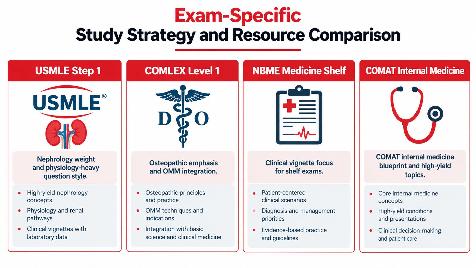
A helpful way to study nephrology is to pair each exam with the kind of reasoning it rewards.
For USMLE Step 1, renal questions often start with physiology and build outward. A board-style vignette might describe changes in effective arterial blood volume, then ask what happens to renin, aldosterone, or fractional sodium excretion. Your job is to move from mechanism to consequence. If you know how the nephron behaves, the answer choices usually narrow quickly.
For COMLEX Level 1, the renal science still matters, but the presentation may feel broader and more integrated. A composite vignette might mix acid-base data, blood pressure findings, and a medication clue. The safest approach is the same one you use in physiology: identify the primary process, localize the problem, then connect it to the clinical picture.
For the NBME Medicine Shelf and COMAT Internal Medicine, nephrology often becomes more bedside-facing. A composite vignette may give you a patient with vomiting, oliguria, muddy brown casts, or heavy proteinuria and ask for the next diagnostic step, the likely category of kidney injury, or the complication that needs treatment first. Here, transporter details matter less than pattern recognition.
One more perspective helps. If you are considering nephrology as a career, interest in the specialty rises and falls over time, as noted earlier from ASN match reporting. That does not change how you should study for exams. Boards reward clear renal reasoning, whether you plan to specialize in it or need to master it now.
Resource comparison without overbuying
Students often collect renal resources the way patients retain sodium. The result is more volume, not better filtration.
A better plan is to assign each resource a job.
- UWorld: Best for practicing integrated renal reasoning and reading detailed explanations after missed questions.
- AMBOSS: Useful when you need a quick explanation of a mechanism, then want to test it right away.
- TrueLearn: Common for students preparing for COMLEX who want familiarity with that exam style.
- COMQUEST: Often chosen for COMAT and broader osteopathic exam practice.
The trap is easy to miss. If you keep opening new banks before finishing review of old mistakes, you are increasing exposure but not improving retrieval. Renal learning works more like interpreting a urinalysis than skimming a textbook. The useful signal comes from a few high-yield findings that you learn to recognize quickly.
| Resource | Strength | Limitation | Best use case |
|---|---|---|---|
| UWorld | Detailed explanations tied to pathophysiology | Slow if you are still shaky on basics | Step-style integration and error review |
| AMBOSS | Fast reference plus question practice | Easy to read passively instead of testing yourself | Filling mechanism gaps during content review |
| TrueLearn | Familiar COMLEX-style question exposure | Explanation style may not fit every learner | COMLEX-focused practice sets |
| COMQUEST | Helpful for COMAT and osteopathic review | Less suited for first-pass teaching of renal physiology | COMAT reinforcement |
A simple rule helps. Use one primary question bank, one secondary reference, and one error log.
For students who want one-on-one support, Ace Med Boards offers tutoring for USMLE, COMLEX, and Shelf preparation. It fits best as a supplement to a primary question bank, not as a substitute for doing and reviewing questions.
A Realistic Six-Week Study Plan Vignette
Composite learner: Sam is a second-year medical student preparing for a medicine-style renal block while juggling a busy schedule. Sam doesn't need a heroic plan. Sam needs a repeatable one.
Weeks 1 through 3
Week 1: Build the scaffold. Draw the nephron by hand. Label where sodium, water, glucose, potassium, and bicarbonate move. Review autoregulation, GFR, and diuretic sites of action.
Week 2: Add acute kidney injury and chronic kidney disease. Do question sets on pre-renal, intrinsic, and post-renal patterns. Write one sentence after each missed question: “The clue I missed was…”
Week 3: Group glomerular disease by pattern, not by long lists. Make a one-page nephritic versus nephrotic comparison sheet. Review active sediment, proteinuria logic, and when a biopsy becomes part of the discussion.
Weeks 4 through 6
Week 4: Drill electrolytes and acid-base every day in short bursts. Rework the same calculations until the order feels automatic.
Week 5: Switch to mixed timed blocks. Review explanations slowly. Keep an error log with three columns: concept missed, why you missed it, and what clue should trigger the right approach next time.
Week 6: Use practice testing and review. Revisit only the highest-yield misses. A crowded plan is less helpful than a consistent one.
Short, repeated renal review usually beats one exhausting weekend binge.
If your study time feels fragmented, a structured medical student study schedule can help you turn these themes into actual calendar blocks.
Key Takeaways and Next Steps for Nephrology Prep
Nephrology becomes much less intimidating once you build it in the right order. Start with filtration and tubule function, then move to AKI versus CKD, then glomerular patterns, then electrolytes and acid-base.
A few priorities are worth keeping front and center:
- Master physiology first: GFR, autoregulation, and nephron segment jobs explain many later questions.
- Think in trends: CKD questions often reward pattern recognition over reaction to one isolated value.
- Read the urine carefully: Sediment and protein clues can localize the problem faster than broad memorization.
- Use a fixed acid-base sequence: pH, primary process, anion gap, compensation.
One historical point also helps explain why the field is so structured. Kidney Disease: Improving Global Outcomes (KDIGO) was created in 2003 after concerns about fragmented nephrology guideline development, and its first major guideline publication, the Hepatitis C Guideline, appeared in April 2008; earlier roots included the 1997 publication of four DOQI guidelines (KDIGO history in Nephrology Dialysis Transplantation). That guideline culture is one reason renal questions often feel systematic. The field expects classification, staging, and consistent reasoning.
Bookmark the physiology, acid-base, and renal review materials you plan to revisit. Then begin each study block by retrieving the concept from memory before you look anything up.
Sources
- Global kidney disease burden analysis in Nephrology Dialysis Transplantation
- KDIGO 2024 CKD Guideline
- KDIGO 2024 summary recommendations on albuminuria confirmation
- American Family Physician review of chronic kidney disease staging
- PubMed review on chronic kidney disease of unexplained cause and CKDx
- American Society of Nephrology fellowship match data
If nephrology still feels scattered after self-study, Ace Med Boards offers diagnostic tutoring and personalized study-plan support for students preparing for USMLE, COMLEX, and Shelf exams. A focused review can help you identify whether your main issue is physiology, question interpretation, or acid-base workflow, then build a plan around that.