
You've reviewed muscles, nerves, and imaging, yet a board-style vignette still leaves you staring at the symptom pattern, unsure which structure has been injured. Human anatomy becomes more manageable when you stop treating it as a catalog of names and instead trace each problem through structure → function → blood or nerve supply → lesion → expected deficit. That sequence turns spatial relationships into a repeatable localization method for USMLE, COMLEX-USA, and clinically oriented anatomy questions.
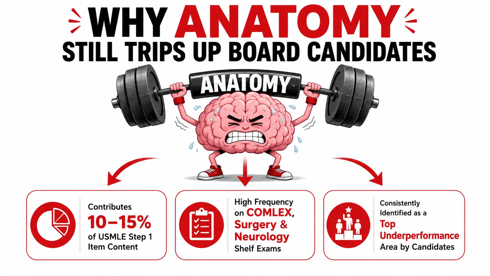
Why Anatomy Still Trips Up Board Candidates
Anatomy questions rarely ask only, “What is this structure?” A vignette may describe a fracture, a sensory change, an abnormal gait, or a cranial nerve finding and expect you to infer the injured structure from the pattern. Image-rich prompts, cross-sectional views, and cadaver-style identification questions test whether you can translate a three-dimensional relationship into a clinical deficit.
The difficulty is often not a lack of effort. Students spend substantial time in the cadaver lab, reviewing diagrams, and memorizing origins, insertions, foramina, and nerve roots. Yet isolated facts can collapse under time pressure when the question changes the patient's position, introduces a vascular injury, or places the lesion proximal to the branch you memorized.
Replace lists with a causal chain
Start with the deficit, then work backward:
- Name the function that has changed. Is the problem motor, sensory, autonomic, or vascular?
- Map the finding to a structure. Use the distribution, not a single buzzword.
- Trace the supply. Identify the nerve, artery, or spinal segment supporting that function.
- Locate the lesion along the pathway. Decide whether it lies at a root, plexus level, named nerve, branch, or target organ.
- Predict one additional finding. A correct localization should explain more than the symptom that brought you there.
This is closely related to the broader habit of connecting findings to mechanisms described in clinical reasoning for medical exams. A compressed textbook table can tell you that a nerve innervates a muscle. A vignette asks whether the lesion occurred before or after a branch left the nerve, and whether the sensory deficit matches the same level.
Practical rule: If your answer explains only one finding, keep localizing. If it explains the motor deficit, sensory territory, reflex, and mechanism of injury, you're probably at the right level.
Build spatial fluency deliberately
Use anatomical planes and directional terms before regional memorization. A learner who can identify what is medial, deep, proximal, or posterior can interpret a new image more effectively than one who remembers a long list without orientation. The aim isn't to memorize every structure equally. It's to recognize relationships that predict what an injury will disrupt.
Historical anatomy shows why this evidence-based approach matters. Mondino de' Liuzzi's Anatomia Corporis Humani, produced in 1316, was described by the U.S. National Library of Medicine as the first modern dissection manual and remained widely used through the 16th century. Andreas Vesalius earned his doctorate at Padua in 1537 and completed De humani corporis fabrica six years later at age 27. The shift from inherited authority toward direct observation created the foundation for modern anatomical education and clinical precision.
Anatomical Planes and Directional Terms in Clinical Context
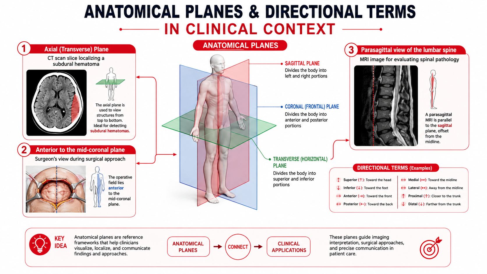
Anatomical language is a coordinate system. It lets you describe where a lesion lies without relying on the reader's viewpoint, which is essential because a CT, MRI, operative field, and physical examination may present the same region differently.
Read the plane before naming the structure
A sagittal plane divides the body into right and left portions. A mid-sagittal image passes through the center, while a parasagittal image lies beside the midline. This is useful for tracing the lumbar spine, spinal cord, ventricles, or a lesion relative to the vertebral canal.
A coronal plane divides anterior from posterior. It can clarify whether a mass is in front of or behind a major compartment, while a transverse plane, also called axial, divides superior from inferior. Axial images are especially useful for following vessels, identifying a subdural collection relative to the falx, and comparing structures at the same level.
An oblique plane cuts at an angle. It often appears when imaging follows a structure that doesn't align neatly with the body's principal planes, such as a joint, a nerve pathway, or an intervertebral space.
Convert bedside language into coordinates
Directional terms become easier when paired with a deficit:
| Clinical phrase | Anatomical translation |
|---|---|
| Wrist drop | Loss of wrist and finger extension, usually pointing toward the radial nerve pathway |
| Foot drop | Weak ankle dorsiflexion, requiring consideration of the common peroneal or deep peroneal pathway, root involvement, or muscle disease |
| Proximal weakness | Greater impairment near the trunk than distally |
| Medial to the artery | The structure lies closer to the body's midline than the artery |
| Deep to the fascia | The structure lies farther from the skin surface |
| Ipsilateral deficit | The deficit occurs on the same side as the lesion |
| Contralateral deficit | The deficit occurs on the opposite side |
Superior and inferior describe position toward or away from the head. Proximal and distal describe position toward or away from a limb's attachment. Superficial and deep describe distance from the surface. In dentistry, proximal surfaces face adjacent teeth, while distal surfaces face away from the midline, showing why directional language remains useful beyond musculoskeletal questions.
Palmar refers to the hand surface, plantar to the foot sole, and dorsal to the posterior surface of the hand or foot. Ventral and anterior often overlap in clinical description, but always follow the convention used by the image or question stem.
Try this translation exercise: “The lesion is deep, lateral, and distal to the elbow, with ipsilateral sensory loss over the dorsal first web space.” Before naming a nerve, identify the coordinates. This habit prepares you to use anatomical terms and directions as localization tools rather than vocabulary to memorize.
Structure to Deficit Applied to the Brachial Plexus
The brachial plexus is a useful model because it forces you to localize along a pathway rather than jump immediately to a named nerve. Its organization is roots → trunks → divisions → cords → terminal branches. The roots are C5 through T1, the upper trunk combines C5 and C6, the middle trunk continues C7, and the lower trunk combines C8 and T1.
A board vignette becomes more tractable when you ask where the pattern first becomes possible. A distal radial nerve injury produces a different combination of weakness and sensory loss from a posterior cord lesion, even though both can affect extension.
Hypothetical one
A patient sustains a fracture at the surgical neck of the humerus. Afterward, the wrist and fingers fall into flexion, and sensation is reduced over the first dorsal web space.
Structure: The radial nerve is vulnerable along the humerus and carries motor fibers to the extensor compartments.
Function: It supports wrist and finger extension.
Supply: Its sensory distribution includes the dorsal first web space.
Lesion: The combined motor and sensory pattern localizes to the radial nerve rather than an isolated root.
Expected deficit: Wrist drop with sensory loss in the characteristic dorsal web-space territory.
The concealed-answer technique makes retrieval active. Cover the final two columns of a table, read the symptom cluster, and state the structure before checking the answer. Then alter one variable. If the same patient has preserved wrist extension but cannot abduct the arm, the lesion no longer fits the radial nerve. You should reconsider the axillary nerve, upper trunk, or a more proximal process based on the new motor pattern.
| Plexus level | Sensory territory | Motor function | Classic lesion |
|---|---|---|---|
| C5 root | Lateral shoulder and upper arm pattern | Shoulder abduction and elbow flexion contributions | Root lesion with weakness in C5-linked movements and altered reflex contribution |
| Upper trunk, C5-C6 | Lateral upper limb pattern | Shoulder abduction, external rotation, and elbow flexion contributions | Erb-Duchenne pattern after traction to the neck and shoulder |
| Middle trunk, C7 | Posterior arm and forearm contributions | Elbow, wrist, and finger extension contributions | C7-predominant weakness and sensory change |
| Lower trunk, C8-T1 | Medial forearm and hand pattern | Intrinsic hand and finger flexion contributions | Lower trunk injury with hand weakness and medial forearm sensory loss |
| Radial nerve | Dorsal radial hand, especially the first web space | Wrist and finger extension | Wrist drop after injury near the humerus |
| Median nerve | Lateral palmar hand and selected fingertips | Thenar opposition and many forearm flexor actions | Thenar weakness, sensory loss in its hand territory, or a hand-of-benediction pattern with proximal injury |
| Ulnar nerve | Medial hand and selected fingers | Interossei and many intrinsic hand actions | Loss of finger abduction and adduction with ulnar-sided sensory change |
Test the level, not just the label
A motorcycle handlebar injury that forces the neck away from the shoulder can injure the upper trunk. The resulting Erb-Duchenne posture reflects loss of C5-C6-dominant functions, including shoulder abduction and external rotation with impaired elbow flexion. That pattern differs from a radial nerve lesion because the deficit begins more proximally and affects a broader group of movements.
A proximal median nerve lesion can produce a hand-of-benediction appearance when the patient attempts to make a fist, along with thenar sensory loss and weakness of forearm muscles supplied before the wrist. A distal median lesion at the carpal tunnel would not reproduce the same proximal motor findings. That single distinction often separates a memorized association from true localization.
For additional timed practice, use neurology Shelf exam practice questions to force the same sequence: symptom, function, supply, lesion level, and predicted deficit.
Lesion Localization Across Cranial Nerves Abdomen Pelvis and Lower Limb
A symptom cluster narrows the field more effectively than a structure list. The concealed-answer column below is designed for retrieval. Read the first two columns, say the likely structure aloud, and only then reveal the answer and confirmatory sign.
| Region | Presenting symptom cluster | Likely structure | Confirmatory sign |
|---|---|---|---|
| Cranial nerve | Ptosis, eye positioned down and out, dilated pupil | Concealed answer: CN III | The eye cannot adduct, elevate, or depress normally, and parasympathetic pupil function is affected |
| Cranial nerve | Inability to abduct the eye with horizontal diplopia | Concealed answer: CN VI | Weak lateral rectus function |
| Cranial nerve | Facial weakness involving the forehead and lower face | Concealed answer: Facial nerve, CN VII | Weakness of facial expression on the affected side |
| Cranial nerve | Hoarseness after a lesion near the larynx | Concealed answer: Recurrent laryngeal nerve | Vocal fold movement is impaired, while the superior laryngeal branch has a different functional territory |
| Abdomen | Poorly localized visceral discomfort that later becomes sharply localized near the parietal peritoneum | Concealed answer: Visceral-to-somatic pain transition | Increasing focal tenderness as parietal peritoneal irritation develops |
| Abdomen | Groin bulge that follows the inguinal canal pathway | Concealed answer: Inguinal hernia | Relationship to the inferior epigastric vessels and inguinal ring helps distinguish the subtype |
| Abdomen | Groin bulge inferior to the inguinal ligament | Concealed answer: Femoral hernia | Location in the femoral canal region |
| Abdomen | Pain with resisted hip flexion or hip extension | Concealed answer: Psoas irritation | A psoas maneuver reproduces symptoms, though clinical interpretation requires context |
| Abdomen | Pain associated with lateral trunk movement | Concealed answer: Quadratus lumborum region | Symptoms change with ipsilateral or contralateral side bending |
| Pelvis | Saddle anesthesia with impaired bladder or bowel control | Concealed answer: Sacral roots, especially S2-S4 pathways | Perineal sensory loss and pelvic floor dysfunction |
| Pelvis | Pelvic floor weakness with altered continence | Concealed answer: Pudendal and S2-S4-related structures | Reduced voluntary pelvic floor contraction |
| Lower limb | Foot drop with sensory change over the dorsum of the foot | Concealed answer: Common peroneal nerve near the fibular neck | Weak dorsiflexion and eversion, with localization refined by inversion testing |
| Lower limb | Numbness over the anterolateral thigh without motor loss | Concealed answer: Lateral femoral cutaneous nerve | Sensory symptoms beneath the inguinal ligament distribution, with preserved quadriceps strength |
| Lower limb | Trendelenburg gait or pelvic drop during single-leg stance | Concealed answer: Superior gluteal nerve or gluteus medius | Weak hip abduction on the unsupported side |
| Lower limb | Hip injury with concern for femoral head perfusion | Concealed answer: Medial circumflex femoral artery | The vascular relationship helps explain why certain hip injuries threaten the femoral head |
Demonstrate the reasoning with one variable change
Suppose a patient has foot drop and difficulty everting the foot after compression near the fibular neck. The common peroneal nerve is a reasonable first localization because it supplies the deep and superficial peroneal divisions. Now change one variable: the patient also has weak inversion and symptoms extending above the knee.
That broader pattern makes an isolated common peroneal lesion less satisfying. Consider a more proximal lesion, such as an L5-related process or a sciatic pathway injury, and use the sensory distribution, reflexes, and additional muscle testing to refine it. The point isn't to force every vignette into one memorized label. It's to ask which lesion explains the greatest number of findings with the fewest exceptions.
Apply the same method to cranial and abdominal anatomy
For a CN III palsy, the structure-function chain is straightforward: the oculomotor nerve supplies most extraocular movements, lifts the eyelid, and carries parasympathetic fibers to the pupil. A down-and-out eye, ptosis, and a dilated pupil therefore point to a lesion affecting both somatic motor and parasympathetic components. A pupil-sparing pattern changes the localization and should prevent an automatic answer.
In the abdomen, distinguish visceral pain from somatic pain before naming an organ. Visceral pain is often poorly localized because it travels with autonomic afferents, while parietal peritoneal irritation produces sharper localization through somatic pathways. The same structure-function-supply logic applies to whether an organ lies intraperitoneally or retroperitoneally, but don't treat those compartments as interchangeable.
Cross-sectional anatomy reinforces this reasoning. A CT image is not a flat collection of labels. Identify the plane, orient anterior and posterior, locate a stable landmark such as the vertebral body, and then follow vessels or organs across adjacent slices. Abdominal CT interpretation guidance can complement this anatomy-first approach, but every image should still be interpreted through relationships and expected deficits.
An Eight Week Anatomy Study Plan for USMLE and COMLEX
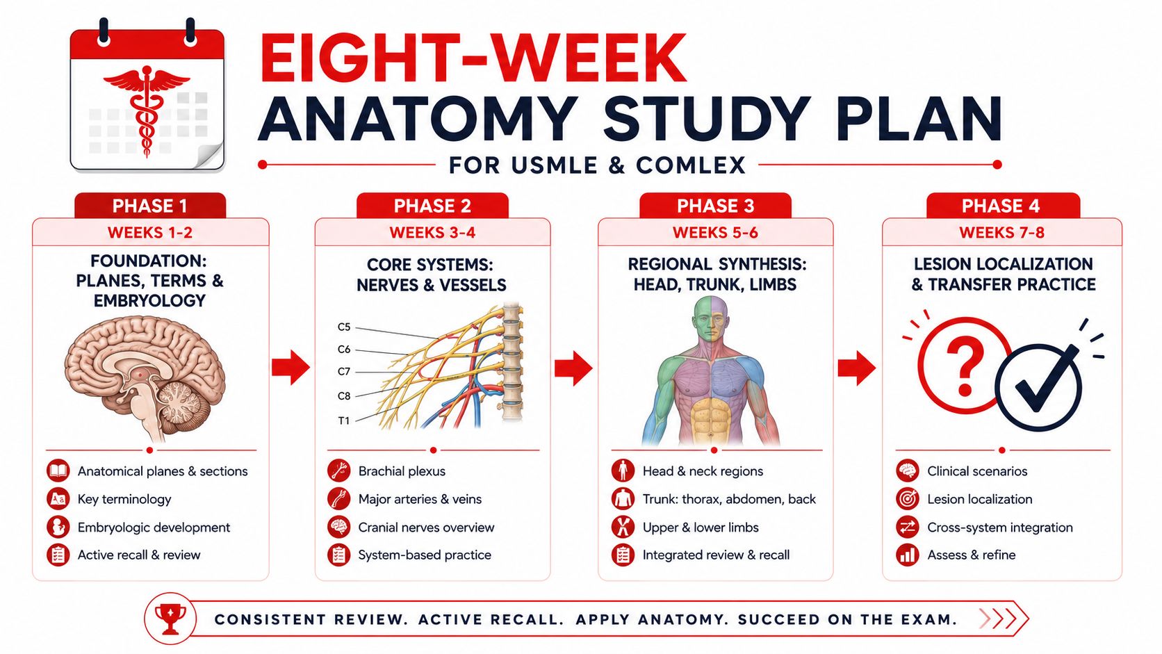
Anatomy improves when each study block produces a retrievable explanation, not another passive reread. This eight-week plan uses four phases, with each phase lasting two weeks. Adjust the daily volume to your full study schedule, but keep the order of skills intact.
Phase one builds orientation
Weeks one and two focus on sagittal, coronal, transverse, and oblique planes, directional terms, body regions, and embryologic organization. Use labeled cross-sections and cadaveric video, but pause before labels appear. State the plane, orientation, and two relationships from memory.
Create short cards for terms that you repeatedly reverse. Each card should include a clinical sentence, such as “the lesion is deep and medial to the vessel,” rather than a dictionary definition alone.
Phase two organizes pathways
Weeks three and four shift to regional anatomy. Group nerves with their roots, motor functions, sensory territories, reflexes, and vulnerable locations. Do the same for major vessels and clinically relevant compartments.
Use the comparison tables as concealed-answer drills. Cover the likely structure and confirmatory sign, read the symptom cluster, and explain why the nearest competing structure is less likely. A card that asks “What does the radial nerve do?” is less powerful than one that asks “Which lesion causes wrist drop with first dorsal web-space sensory loss?”
Phase three connects anatomy to questions
Weeks five and six integrate anatomy-tagged question-bank items with regional review. The assigned schedule calls for at least 25 anatomy-tagged items per day, but that number should be treated as a planning target rather than a universal requirement. If careful review takes longer, reduce volume and preserve the explanation.
For every missed or guessed item, write five margin prompts:
- Structure: What was injured or being tested?
- Function: What movement, sensation, or physiologic role changed?
- Supply: Which nerve, artery, root, or compartment is involved?
- Lesion: Where along the pathway is the damage?
- Deficit: What second finding should appear?
Tag errors by type, such as plane confusion, branch confusion, sensory-map error, or lesion-level error. Improvement should be judged by fewer repeated error tags, stronger timed accuracy, and the ability to explain why distractors fail. Don't judge the method by an undocumented score promise.
Phase four trains transfer
Weeks seven and eight connect chest radiographs, abdominal CT, and brain MRI orientation to cadaveric plates and regional diagrams. During the final week, use full-length NBME practice according to the current official rules and materials available to you, then review anatomy errors by region and error tag.
A practical weekly rhythm might look like this:
| Study block | Primary task | Output |
|---|---|---|
| First block | Active recall of pathways | One completed structure-to-deficit map |
| Second block | Question practice | Error tag and distractor explanation |
| Third block | Image orientation | Plane, landmarks, and relationships |
| Final review | Spaced repetition | Recalled answers before card reveals |
The USMLE Step 1 study plan can sit alongside the official USMLE Step 1 Content Outline, which should control the exam scope and terminology. COMLEX learners should also consult current NBOME materials for the relevant blueprint and format rather than assuming the two examinations are identical.
Common Pitfalls Ethics Gaps and Next Steps for Learners
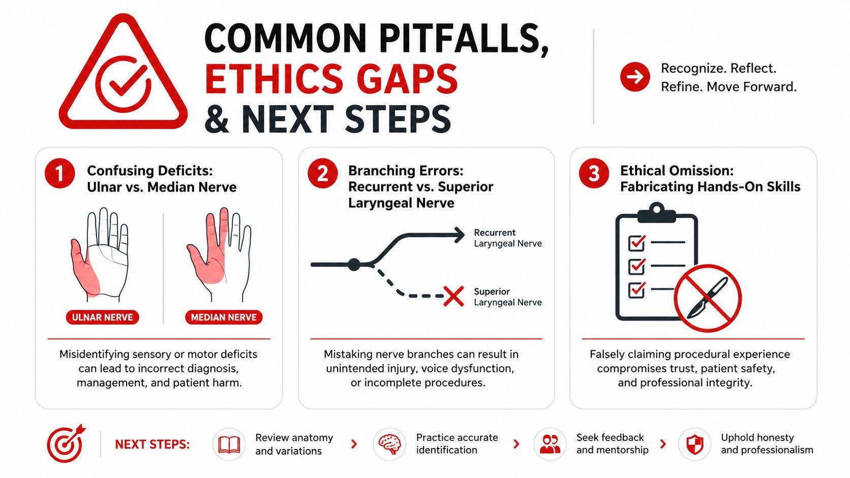
The most persistent musculoskeletal error is confusing a deficit with a nerve name before checking the distribution. Ulnar and median lesions can both produce hand weakness, but their sensory territories, intrinsic muscle findings, and effects on finger positioning differ. Similarly, recurrent laryngeal and superior laryngeal branches should be separated by function and course, not by the shared word “laryngeal.”
Retroperitoneal and intraperitoneal relationships create another frequent trap. When a question describes pain, hemorrhage, or surgical access, compartment location changes the expected spread and neighboring structures. Ask where the organ lies, what surrounds it, and which vessels or nerves pass nearby.
Evaluate digital anatomy resources ethically
Visual polish doesn't guarantee trustworthy sourcing. A 2024 PubMed study analyzing 3,883 anatomy images found that 81.2% of codable skin tones were light, 61.6% of codable sex-binary images depicted males, and, where body size or age was identifiable, 85.0% were adults. Read those findings in the PubMed study on representation in anatomy imagery, and ask whether a resource distinguishes common anatomy from commonly illustrated anatomy.
Provenance and consent deserve the same scrutiny. A 2026 PubMed study found that only 21.3% of 239 anatomy videos disclosed provenance and 4.2% documented multimedia-specific consent. The PubMed research on provenance and consent in anatomy videos supports a practical checklist:
- Provenance: Can the resource identify where the specimen, image, or model came from?
- Consent: Does it explain whether educational multimedia use was authorized?
- Representation: Does it show meaningful variation in age, sex, body size, and skin tone?
- Transparency: Does it distinguish donor-based material from synthetic or AI-generated illustrations?
- Confidentiality: Does it avoid identifiable patient information and misleading claims of hands-on experience?
AI-generated illustrations can clarify concepts when donor material isn't available, but they shouldn't be presented as anatomically authoritative without labeling and verification. For exam preparation, use them as explanatory supplements, then confirm relationships in established references and official materials.
Human anatomy is also a moving framework. Adults are commonly taught as having 206 bones and about 9 major organ systems, although classifications can describe 10 to 12 systems; organ counts are often given as 78, with clinical discussions ranging from 78 to 80, and one Gray's Anatomy classification reached 79 after recognition of the mesentery as a true organ in the 2020 printing of its 42nd edition (clinical anatomy overview). The adult vertebral column is commonly organized as 33 vertebrae, with 7 cervical, 12 thoracic, 5 lumbar, 5 sacral, and 4 coccygeal segments (vertebral column reference). These standards help you communicate, while classification changes remind you that anatomy is evidence-based rather than frozen.
For individualized support, use the completed comparison table to identify recurring weak spots. One-on-one USMLE or COMLEX tutoring can then focus on explaining distractors, interpreting images, and rehearsing lesion localization rather than repeating an entire textbook. This is educational content for examination preparation, not individualized medical advice or patient treatment guidance.
Ace Med Boards offers personalized one-on-one tutoring and question-analysis support for USMLE and COMLEX preparation, including anatomy-focused work on structure-function relationships and lesion localization. If you want help reviewing your error tags and building a targeted study plan, visit Ace Med Boards to explore a free consultation without treating tutoring as a guarantee of any particular score or outcome.
Sources
- U.S. National Library of Medicine article on the history of anatomical dissection and publishing
- NCBI review of skeletal muscle fiber types and function
- PubMed research on muscle fiber mechanics and recruitment
- PubMed study on representation in anatomy images
- PubMed study on provenance and consent in anatomy videos
- USMLE Step 1 Content Outline