
You're staring at an anatomy question or a radiology image, and the words all look familiar until one term flips the meaning. That's usually where readers lose points, not because they forgot the structure, but because they misread anatomical terms and directions. The fix is a standardized vocabulary tied to anatomical position, then a steady habit of translating each term into what the test writer wants.
Why Anatomical Terms and Directions Matter on Exam Day
A common exam-day miss goes like this. A student sees a stem about abdominal pain, notices a structure described as “lateral,” and answers too quickly because the word sounds simple. Then the diagnosis hinges on whether the finding was medial or lateral, or whether it was anterior rather than posterior.
That matters because anatomical terms and directions are not loose descriptors. They are fixed reference terms built from standard anatomical position, and that consistency is what keeps anatomy, radiology, and physical-exam language readable across settings. The first widely recognized standardization effort came with the Basle Nomina Anatomica in 1895, which deliberately reduced naming chaos by choosing about 5,000 terms from roughly 50,000 competing names (Nomina Anatomica). Later, Terminologia Anatomica became the international standard in 1998, reflecting the broader push toward coordinated anatomical language rather than local custom (Terminologia Anatomica history).
What the exam tests is often not raw recall of anatomy, but whether you can place a structure correctly in space. A mass can be medial to one landmark and lateral to another, or a lesion can be superficial instead of deep. A single swapped word can flip your interpretation.
Practical rule: When a question uses directional language, slow down and ask, “Relative to what reference point?”
A calm review of the vocabulary pays off quickly because the same terms keep appearing across USMLE, COMLEX-USA, and Shelf-style vignettes. The sections below build the system step by step, then show how the test writer uses it to mislead you.
Anatomical Position and the Core Directional Pairs
Anatomical position is the reference posture that makes direction words mean the same thing for everyone. The person stands upright, faces forward, keeps the arms at the sides, and turns the palms forward. In that posture, anterior and posterior stay anchored to the body's front and back, not to where the viewer happens to stand (positional and directional terms).
The directional pairs that do the heavy lifting
- Anterior and posterior: Anterior means toward the front of the body, and posterior means toward the back. The sternum is anterior to the heart, and the spine is posterior to the sternum.
- Superior and inferior: Superior means toward the head, and inferior means toward the feet. The nose is superior to the mouth, and the umbilicus is inferior to the sternum.
- Medial and lateral: Medial means closer to the midline, and lateral means farther away from it. The sternum is medial to the shoulders, while the shoulders are lateral to the sternum.
- Proximal and distal: These terms are for appendages, not the trunk. Proximal means nearer the trunk or point of attachment, and distal means farther away. The elbow is proximal to the wrist, and the fingers are distal to the elbow (Directional Terms/01:_Introduction_to_Anatomy_and_Anatomical_Terms/1.03:_Directional_Terms)).
- Superficial and deep: Superficial means closer to the body surface, and deep means farther from it. The skin is superficial to muscle, and the heart is deep to the ribs.
Cranial and caudal show up often as synonyms for superior and inferior, especially in embryology and imaging language. If you keep the anatomical position in mind, the synonym set becomes much easier to read.
A useful habit is to attach each word to a landmark. The great toe is distal to the ankle, the umbilicus is medial to the flanks, and the nose is superior to the mouth. Once those landmarks feel automatic, the rest of the terminology becomes less like memorization and more like translation.
The reference posture is the key, not the observer's point of view.
Body Planes and Axes in Clinical Imaging
Body planes turn the directional language into visible slices. They help you read CT, MRI, and anatomy diagrams without guessing which side of the body you're looking at. For a broader exam-style orientation review, a comprehensive head to toe assessment guide can be a useful companion when you're connecting surface landmarks to imaging and bedside exam language.
| Plane | Divides | Perpendicular Axis | Common Imaging | Example View |
|---|---|---|---|---|
| Sagittal | Left from right | Mediolateral | Brain MRI, sagittal spine MRI | Shows the body from the side, often useful for midline structures |
| Coronal, or frontal | Anterior from posterior | Anteroposterior | Coronal CT face, coronal MRI | Shows the body as if split into front and back portions |
| Transverse, or axial, or horizontal | Superior from inferior | Superoinferior | CT abdomen, axial brain CT | Shows a cross-section, often used in emergency imaging |
| Oblique | Angled off the standard planes | Varies with the tilt | Cardiac imaging, musculoskeletal imaging | Shows a structure along a slanted path |
The main job of the plane is to tell you what kind of slice you're seeing. A sagittal image divides left from right, so it's useful when a question describes a structure near the midline. A coronal image divides anterior from posterior, which is why it often helps with face, chest, and abdominal relationships. A transverse slice divides superior from inferior, so it's the classic cross-sectional view on many CT studies.
Clinical habit: Name the plane first, then read the direction terms inside that plane.
If you want to practice that skill on thoracic imaging, the chest X-ray interpretation guide is a natural next stop. It helps connect what you see on a film to the directional language used in stems and reports.
Paired Terms That Students Confuse Most Often
Some pairs look interchangeable until the question punishes that assumption. The safest way to learn them is to rewrite the same sentence with one word swapped and notice how the anatomy changes. That's how the exam writer uses language, too.
| Pair | Definition | Clinical Example Sentence |
|---|---|---|
| Anterior vs posterior | Front vs back of the body | The sternum is anterior to the heart, while the spine is posterior to it. |
| Superior vs inferior | Toward the head vs toward the feet | The diaphragm is superior to the liver, and the bladder is inferior to the umbilicus. |
| Medial vs lateral | Toward the midline vs away from it | A medial meniscus tear affects the inner side of the knee, while a lateral tear affects the outer side. |
| Proximal vs distal | Nearer vs farther from the point of attachment, limbs only | The elbow is proximal to the wrist, and the fingers are distal to the elbow. |
| Superficial vs deep | Near the surface vs farther from the surface | The skin lesion is superficial, but the abscess lies deep to the abdominal wall. |
The medial and lateral pair trips students because both sound like “side.” The test, though, is about side relative to the midline. If a stem changes one word, the entire structure can change.
The proximal/distal pair is even more specific. It applies to limbs and limb-like structures, not to the trunk. So a stem should not ask whether the esophagus is proximal or distal in the same way it asks whether the forearm is proximal or distal. For a quick terminology refresh that stays close to the language used in medical training, the medical terms and terminology reference can help anchor the word choices you see in class and on exams.
For most board questions, the safest move is to translate the phrase into plain English before you answer. If the sentence says “the lesion is lateral,” ask yourself, lateral to what? If it says “the pain is distal,” confirm that the structure involved is a limb.
Labeled Orientation Examples You Will See on Vignettes

A chest X-ray often looks reversed at first glance because you're not looking at the patient from the patient's own point of view. On a PA chest X-ray, the image is typically read as if you're facing the patient, so the patient's right appears on the viewer's left, and the patient's left appears on the viewer's right. That's why the heart silhouette can seem to sit on the viewer's right side while still being on the patient's left.
On an axial abdominal CT, the reader usually imagines looking up from the feet. That orientation keeps the patient's right and left consistent with the anatomy, even though the slice may feel backward at first. It's a common place for students to miss a laterality clue, especially when a stem shifts from a physical exam description to an imaging description.
A supine patient adds another layer. In that position, the patient lies on the back, but anterior still means toward the front of the body and posterior still means toward the back. The body position changes, the directional terms do not.
A brain MRI can also test localization in a quiet, indirect way. If a stem refers to a lesion in the frontal lobe on a coronal image, the writer may be checking whether you can map the plane to the lobe without confusing it with the parietal, temporal, or occipital region. That's really a directional-language question dressed up as anatomy.
If you want a second imaging comparison point, the abdominal CT reading guide can be useful for practicing how orientation shifts between the written stem and the image itself.
High-Yield Exam Traps and Common Misconceptions
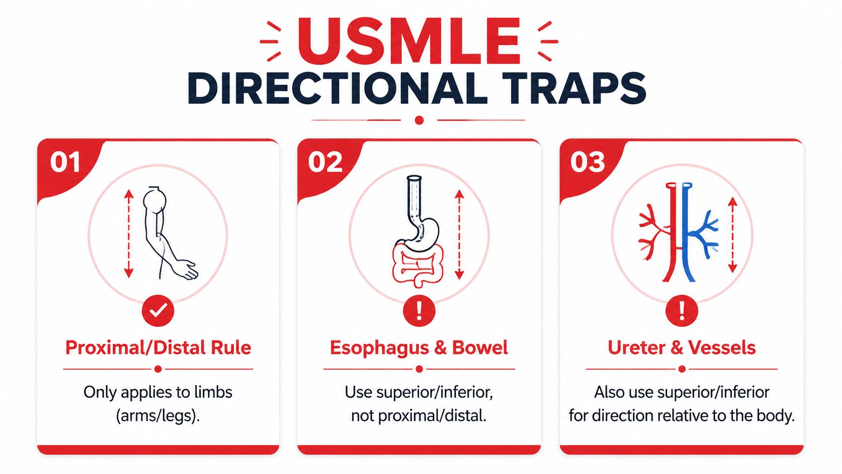
The first trap is treating proximal and distal as if they apply everywhere. They do not. On boards, these words are for limbs and limb-like structures, so they fit the arm, leg, and similar pathways, but not the trunk in the same way. For internal tubes, exam writers usually expect you to use superior and inferior instead.
The second trap is mixing up ventral and dorsal. In human anatomy, ventral lines up with anterior, while dorsal lines up with posterior. Students sometimes carry over veterinary or comparative-anatomy habits and miss the human convention.
The third trap is the gap between official terminology and the language people use in clinic. A review of anatomical terminology notes that students often struggle to translate official terms into clinical language and synonyms, and that this mismatch can lead to mechanical memorization instead of real understanding (European Journal of Anatomy review). Another line of research points to confusion in sex-and-reproductive anatomy, including incorrect positioning and naming errors, which shows that knowing the vocabulary is not always the same as using it correctly in context (conference abstract on sex and reproductive anatomy misconceptions).
The last trap is assuming position words reverse when the patient changes posture. They don't. A prone patient is face down, a supine patient is face up, but anterior and posterior still refer to the body, not the bed. That's why radiology stems sometimes feel like a logic puzzle if you read them from the wrong frame of reference.
Quick filter: If a term feels “off,” check whether you're using the body's reference system or the viewer's.
For item-writing patterns that hinge on traps like these, distractor analysis is a useful way to think about why a tempting wrong answer feels right at first glance.
Quick Self-Test With Original Practice Questions
Use these the way you'd use a short timed block. Read the stem once, pick an answer, then justify it out loud before checking the rationale. If you want a larger bank of original drills, the USMLE Step 1 practice questions page is a natural follow-up.
A chest X-ray report says the cardiac silhouette is shifted toward the patient's left hemithorax. Which term best describes the heart relative to the right lung?
A. Medial
B. Lateral
C. Proximal
D. Distal
Answer: A. Medial. The heart lies closer to the midline than the right lung. Proximal and distal do not fit here because this is not a limb.An axial CT of the abdomen shows an inflamed appendix near the lower right abdomen. Which laterality is most correct for the lesion?
A. Left-sided
B. Right-sided
C. Contralateral to the cecum
D. Superior to the liver
Answer: B. Right-sided. The appendix sits in the patient's right lower quadrant. The trap is reading the image from your own point of view instead of the patient's.On physical exam, a patient has pain at the wrist after a fall. Which structure is proximal to the wrist?
A. Hand
B. Fingers
C. Elbow
D. Thumb tip
Answer: C. Elbow. Proximal means closer to the trunk along a limb. The hand and fingers are distal to the wrist.A question stem describes a skin lesion on the forearm that is close to the surface, not beneath the fascia. Which term fits best?
A. Deep
B. Superficial
C. Posterior
D. Caudal
Answer: B. Superficial. This is a depth question, not a front-back or head-to-foot question.A patient has pain in the left ankle after a right knee injury. Which term describes the relationship between the painful ankle and the injured knee?
A. Ipsilateral
B. Contralateral
C. Medial
D. Proximal
Answer: B. Contralateral. The ankle pain is on the opposite side from the right knee injury. Ipsilateral would mean same side.A stem says a mass lies on the ventral surface of the body. In human anatomy, what does that most nearly mean?
A. Posterior
B. Anterior
C. Distal
D. Lateral
Answer: B. Anterior. In human terminology, ventral corresponds to the front of the body.
Key Takeaways and a Study Plan for Lasting Recall
Anatomical terms and directions only make sense when you anchor them to anatomical position. That's the reference frame the exams assume, whether the question uses anatomy lab language, imaging language, or bedside shorthand.
Three rules deserve extra attention. Proximal and distal apply to limbs, superficial and deep describe depth, and ipsilateral and contralateral describe same-side versus opposite-side relationships. The harder part is not learning the words, but staying alert when clinical language drifts away from textbook phrasing.
A simple study plan helps the terms stick:
- Day 1, pair the opposites: Review anterior/posterior, superior/inferior, medial/lateral, proximal/distal, and superficial/deep in one sitting.
- Day 2, narrate images aloud: Look at a CT or MRI and say the direction terms out loud before checking the label.
- Day 3, annotate an atlas: Write the paired terms on printed anatomy images so the spatial relationship becomes visual.
- Day 7, run a short quiz: Use five stems that mix imaging, bedside exam, and limb localization.
The goal is not memorizing a list. It's training your eye to translate the stem into a map. If you want live drill practice and personalized feedback on weak spots, Ace Med Boards offers diagnostic tutoring and study-plan support for anatomy-heavy board prep, including direction and localization work that often decides whether a question feels easy or tricky.
Ace Med Boards can help you tighten the exact directional skills that show up on USMLE, COMLEX-USA, and Shelf-style questions, especially when the stem mixes imaging, anatomy, and laterality. If you want a calm, targeted review of what's confusing you, visit Ace Med Boards to explore diagnostic tutoring and personalized study-plan support.