
You've reviewed bacterial shapes, toxins, stains, and antibiotic mechanisms, yet a microbiology question still feels like a pile of unrelated facts. That happens because USMLE Step 1 questions rarely test organism names in isolation. They ask you to connect a clinical syndrome, a microbial structure or virulence factor, a laboratory clue, and an antimicrobial target.
The most reliable fix is to study microbiology as a diagnostic reasoning chain: classify the organism, connect it to the disease mechanism, then apply the appropriate diagnostic and treatment logic. This guide uses that chain for USMLE Step 1 preparation, while also identifying distinctions that matter to osteopathic medical students preparing for COMLEX Level 1.
Why Microbiology Feels Overwhelming and How to Fix It
A student may spend an evening memorizing organisms, wake up the next morning, and still miss a question about pneumonia, meningitis, or diarrhea. The problem usually isn't effort. Microbiology asks you to hold several layers in working memory at once: taxonomy, morphology, transmission, virulence, clinical presentation, testing, and pharmacology.
A better approach is to treat each organism like a suspect in a clinical investigation. First, establish its broad identity. Is it Gram-positive, Gram-negative, acid-fast, fungal, viral, or parasitic? Next, ask how it causes disease. Does it adhere, invade tissue, evade immunity, form a biofilm, or produce a toxin? Finally, use the syndrome and laboratory findings to choose the most defensible answer.
Practical rule: Don't memorize a pathogen until you can connect its structure to its clinical behavior.
Historical milestones show why these connections matter. Louis Pasteur's work in 1857 demonstrated that microorganisms drive fermentation, following Antonie van Leeuwenhoek's identification of microorganisms in 1677 and preceding Robert Koch's 1876 work formalizing postulates for linking a specific microbe to a specific disease (historical overview of microbiology). Later milestones, including the identification of Mycobacterium tuberculosis as the cause of tuberculosis and the development of the Gram stain, made microbial diagnosis more systematic.
The classify, connect, and apply method
Use three passes for every new organism:
- Classify: Identify the cell type, stain, morphology, genome pattern, or life-cycle category.
- Connect: Link the organism to its transmission route, virulence factor, tissue preference, and clinical syndrome.
- Apply: Select the confirmatory test and antimicrobial class by asking what structure or process the treatment targets.
This method also makes review more efficient. You can borrow a general principle from effective study systems, namely that organizing information into a repeatable structure improves retrieval, much as creators organize content around recognizable patterns in ViewsMax channel growth advice. For microbiology, the pattern isn't a content format. It's organism, mechanism, clue, test, drug.
Use active recall for medical studying rather than rereading organism lists. Cover the diagnostic-test column, predict the test, and then explain why it fits. If you can recall only the name but not the mechanism, the fact isn't exam-ready.
Key takeaways
- Start with classification, especially the Gram stain or an atypical-organism clue.
- Translate virulence into symptoms, such as toxin-mediated diarrhea or capsule-mediated immune evasion.
- Choose tests by organism biology, not by memorized test lists.
- Choose antimicrobials by target and resistance mechanism, not by syndrome alone.
- Review missed questions by reasoning error, not only by pathogen name.
Classifying Major Pathogen Groups for Board Exams
Classification gives you the first branch point in a vignette. The broad hierarchy is domain, cellular organization, structural class, and species. For board questions, you usually don't need to recite every taxonomic rank. You need to recognize the biological category that explains the presentation.
Begin with the bacterial cell wall
The Gram stain separates bacteria by cell wall structure. Gram-positive organisms retain crystal violet because they have a thicker peptidoglycan-rich wall. Gram-negative organisms lose the primary stain during decolorization and appear pink or red after counterstaining (NCBI explanation of Gram staining).
The result isn't just a color label. It depends on the interaction among crystal violet, iodine, the alcohol decolorizer, and the cell wall. A standard sequence uses crystal violet for about 60 seconds, Gram's iodine for about 60 seconds, 95% alcohol for roughly 10 seconds, and safranin for about 45 seconds (Gram stain procedure). Over-decolorization or under-decolorization can produce a misleading interpretation.
| Group | Defining clue | Board-exam use |
|---|---|---|
| Gram-positive bacteria | Thick peptidoglycan wall, purple after Gram staining | Think about cocci or rods, capsules, spores, and exotoxins |
| Gram-negative bacteria | Outer membrane, lipopolysaccharide, pink or red after counterstaining | Consider endotoxin, pili, capsules, and periplasmic beta-lactamases |
| Mycobacteria | Lipid-rich, mycolic-acid-containing wall | Use acid-fast staining rather than relying on Gram stain |
| Spirochetes | Thin, flexible, spiral-shaped bacteria | Dark-field or specialized testing may be needed |
| Fungi | Eukaryotic organisms with chitin-containing cell walls | Use morphology, culture, antigen, or molecular testing |
| Viruses | Acellular infectious particles requiring host-cell machinery | Classify by genome, envelope, replication strategy, and tropism |
| Parasites | Eukaryotic organisms with complex life cycles | Identify eggs, cysts, larvae, trophozoites, or adult forms |
Mycobacteria and spirochetes are common sources of confusion because they don't behave like typical Gram-stained bacteria. Fungi and parasites require a different mental model entirely, since morphology and life cycle often matter more than bacterial cell-wall architecture. Viruses are best approached through genome type, envelope status, replication location, and characteristic clinical patterns.
Use morphology as a second branch point
After the stain, ask whether the organism is a coccus, rod, curved rod, branching filament, yeast, mold, or intracellular pathogen. Shape alone rarely identifies the organism, but it narrows the field and helps you interpret the next clue.
A Gram-positive coccus in clusters points toward a different set of possibilities than a Gram-negative diplococcus. A branching, weakly acid-fast filament suggests a different diagnostic path than a nonbranching rod. The board-style question often supplies one structural clue and expects you to combine it with the host, exposure, and syndrome.
Pathogenesis and Virulence Factors That Drive Clinical Disease
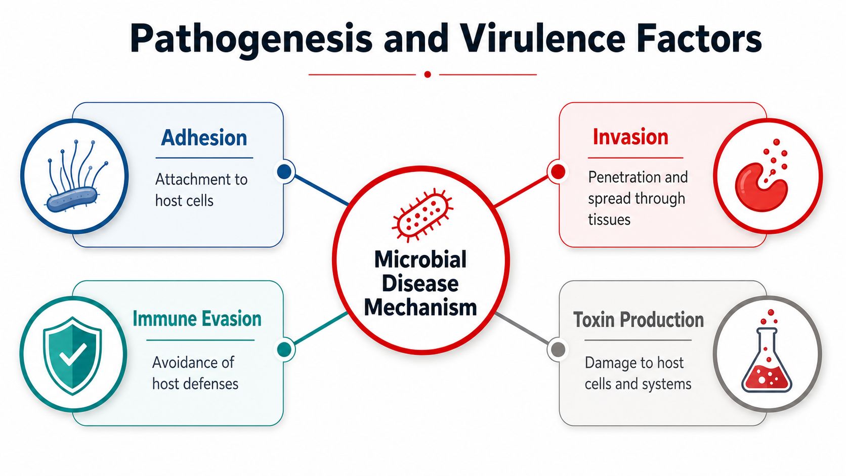
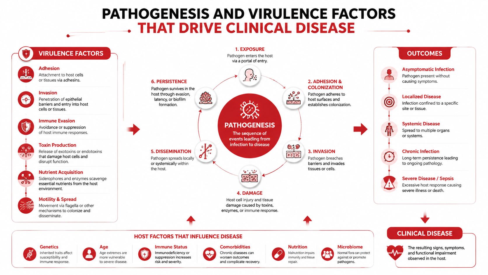
A pathogen's virulence factors explain what happens after exposure. They answer four practical questions: How does the organism attach? How does it enter tissue? How does it avoid host defenses? What damage does it cause?
Adhesion comes first for many infections. Pili, fimbriae, surface proteins, and biofilm-associated structures help organisms remain attached to mucosa, skin, or implanted material. If a vignette emphasizes colonization of a device or persistent infection despite therapy, think beyond simple planktonic growth.
Invasion describes movement through tissue or entry into cells. Enzymes can break down extracellular barriers, while specialized secretion systems can alter host-cell behavior. Intracellular survival then creates a diagnostic clue: the immune response and antimicrobial choice may differ because the organism occupies a protected compartment.
Immune evasion and toxins
Capsules make phagocytosis more difficult. Antigenic variation changes the microbial surface over time. Some pathogens survive inside macrophages or interfere with complement, allowing them to persist despite an intact immune system.
Exotoxins are secreted microbial proteins with specific cellular targets. They may block protein synthesis, disrupt neurotransmitter release, alter intracellular signaling, or damage membranes. Endotoxin, by contrast, is associated with the lipopolysaccharide component of Gram-negative outer membranes. Its effects are driven by host inflammatory signaling and can produce fever, hypotension, and systemic inflammation.
| Mechanism | What it accomplishes | How it appears in a vignette |
|---|---|---|
| Adhesion | Anchors the organism to a host surface | Recurrent mucosal colonization or device attachment |
| Invasion | Enables tissue penetration or intracellular entry | Tissue destruction, abscess formation, or intracellular organisms |
| Immune evasion | Limits phagocytosis or immune recognition | Severe infection in an otherwise intact host or prolonged persistence |
| Toxin production | Produces targeted or systemic cellular injury | Neurologic findings, secretory diarrhea, necrosis, or shock |
| Biofilm formation | Creates a structured community with altered drug tolerance | Chronic catheter, prosthetic, or hardware-associated infection |
Biofilms change the treatment problem
A biofilm is not merely a layer of bacteria hiding from an antibiotic. The extracellular polymeric substance matrix can limit penetration, slow bacterial growth, and support persister cells. Bacteria in biofilms may require 10 to 1000 times higher antibiotic concentrations than genetically equivalent planktonic cells to achieve similar killing (biofilm antimicrobial tolerance).
The biofilm environment also brings cells into close contact, increasing horizontal gene transfer through mechanisms such as plasmid-mediated conjugation and phage-mediated transduction. That makes biofilms reservoirs where antimicrobial-resistance genes can spread within and between species (biofilms and antimicrobial resistance).
For exams, connect biofilm to persistence, foreign material, and treatment difficulty. For clinical education, remember that this is an exam-focused simplification, not a substitute for patient-specific infectious-disease decision-making.
Bugs Versus Clues Table for Rapid Organism Recognition
A useful table doesn't replace reasoning. It gives you a compact set of associations that you can test under time pressure. Read the clinical clue first, predict the organism, then verify the virulence factor and diagnostic method.
| Organism | Key virulence factor | Clinical clue | Diagnostic test |
|---|---|---|---|
| Staphylococcus aureus | Protein A, coagulase, toxins | Abscesses, invasive disease, or toxin-mediated illness | Gram-positive cocci in clusters, catalase positive, coagulase testing |
| Streptococcus pyogenes | M protein, streptolysins | Pharyngitis, skin infection, or postinfectious immune disease | Gram-positive cocci in chains, beta-hemolysis, antigen or culture testing |
| Streptococcus pneumoniae | Polysaccharide capsule | Lobar pneumonia, meningitis, otitis, or sinusitis | Lancet-shaped diplococci, alpha-hemolysis, optochin susceptibility |
| Neisseria meningitidis | Capsule and endotoxin | Meningitis with petechial or purpuric findings | Gram-negative diplococci, culture or molecular testing |
| Neisseria gonorrhoeae | Pili and antigenic variation | Urethritis, cervicitis, pelvic infection, or arthritis | Intracellular Gram-negative diplococci or nucleic acid amplification |
| Corynebacterium diphtheriae | Inhibition of protein synthesis by exotoxin | Pharyngeal pseudomembrane and systemic toxin effects | Culture with toxin testing |
| Clostridium botulinum | Neurotoxin blocking acetylcholine release | Descending flaccid paralysis | Toxin detection and organism testing |
| Mycobacterium tuberculosis | Intracellular survival and lipid-rich wall | Chronic pulmonary symptoms, systemic findings, or granulomas | Acid-fast stain, culture, and nucleic acid testing |
| Candida albicans | Adhesion and biofilm formation | Mucosal disease or opportunistic invasive infection | Budding yeast, pseudohyphae, culture or antigen testing |
| Giardia duodenalis | Adhesion to small-intestinal mucosa | Foul-smelling diarrhea after exposure to contaminated water | Stool antigen, molecular testing, or ova-and-parasite evaluation |
Turn the table into retrieval practice
Don't reread every row repeatedly. Cover the Organism column and infer the bug from the clinical clue. Then cover the Diagnostic Test column and name the test before revealing the answer. A digital deck or printable set of cards can support this process, and microbiology flashcards can serve as one study format among question banks, lecture notes, and official educational resources.
A second pass should compare close alternatives. For example, ask why a capsule matters for one organism, why a toxin matters for another, and why a particular stain is required for a third. That comparison builds flexible recognition instead of brittle memorization.
Antimicrobial Classes and Mechanisms of Action
Antimicrobial questions become easier when you stop treating drugs as long lists. Begin with the microbial structure or process that must be disrupted, then identify the drug class that targets it.
Match the drug to its target
| Target | Major antimicrobial groups | Core mechanism |
|---|---|---|
| Cell wall synthesis | Beta-lactams, glycopeptides | Disrupt peptidoglycan construction or cross-linking |
| Bacterial ribosomes | Aminoglycosides, tetracyclines, macrolides, clindamycin, oxazolidinones | Alter translation or prevent protein synthesis |
| Nucleic acid synthesis | Fluoroquinolones, rifamycins, nitroimidazoles | Interfere with DNA replication or RNA transcription |
| Folate metabolism | Sulfonamides, trimethoprim | Block sequential steps in folate-dependent synthesis |
| Cell membrane integrity | Selected membrane-active agents | Disrupt membrane structure and permeability |
The Gram stain can guide the initial branch, but it doesn't determine the complete treatment plan. Gram-positive and Gram-negative organisms differ in envelope architecture, and the outer membrane of Gram-negative bacteria can limit access to certain targets. A question may therefore ask you to combine stain, site of infection, host factors, and resistance clues.
Board-style translation: Identify the organism class first. Identify the vulnerable target second. Check the resistance mechanism third.
Resistance is a mechanism question
Beta-lactamase production can destroy beta-lactam drugs. Altered penicillin-binding proteins reduce drug binding. Efflux pumps lower intracellular concentrations, while ribosomal modification can prevent protein-synthesis inhibitors from attaching effectively.
Biofilm-associated tolerance adds another layer because reduced growth and poor drug penetration can make genetically susceptible organisms behave differently from free-living cells. On a question, distinguish resistance, which often involves a heritable mechanism, from tolerance, which can reflect the biofilm environment and altered physiology.
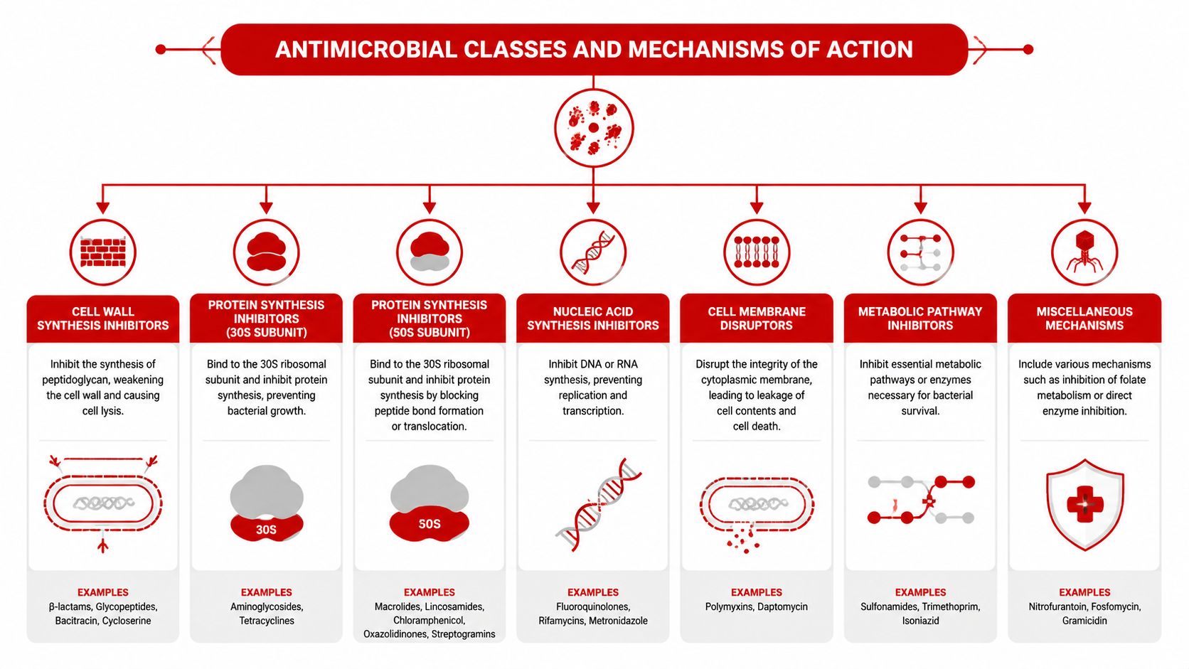
A visual review can help you map drug classes to bacterial structures, but use it actively. Before looking at the diagram, sketch the bacterial cell envelope and place each class at its target. For broader pharmacology integration, pair this exercise with high-yield pharmacology review for USMLE.
Finally, avoid choosing a drug from a symptom alone. “Pneumonia” is a syndrome, not an organism. The stem's age, immune status, exposure, Gram stain, resistance clue, and disease location narrow the answer more reliably than the diagnosis label by itself.
A Worked Framework for Analyzing Microbiology Questions
A microbiology vignette becomes manageable when you read it in a fixed order. Don't begin with the answer choices. First extract the facts that change the probability of each organism.
Four passes through the stem
- Define the syndrome. Decide whether the presentation is primarily respiratory, gastrointestinal, neurologic, genitourinary, skin and soft tissue, bloodstream, or systemic.
- Identify the organism category. Use Gram reaction, morphology, acid-fast behavior, fungal form, viral genome clues, or parasite life-cycle findings.
- Find the mechanism. Look for a capsule, toxin effect, intracellular location, tissue invasion, biofilm, or characteristic transmission route.
- Apply the laboratory and treatment logic. Choose the test that directly detects the suspected organism and the drug class that targets its relevant structure, while accounting for resistance.
Composite example
Composite educational illustration, not a real patient or recalled examination item: An adult develops persistent infection involving implanted hardware. Cultures identify bacteria attached to the device, and standard antimicrobial therapy produces limited clearance. The question asks why the organism is difficult to eradicate.
Start with the syndrome: this is a chronic device-associated infection. Next, identify the mechanism: surface-associated growth suggests a biofilm, not free-floating bacteria. The relevant explanation is the extracellular polymeric substance matrix, slowed growth, and persister-cell formation, all of which can reduce antimicrobial killing.
The best answer should therefore describe altered microbial physiology and matrix-associated tolerance. An answer focused only on a toxin, a capsule, or rapid bloodstream multiplication misses the key clue. The treatment question that follows would require a separate evaluation of organism identity, susceptibility testing, source control, and the clinical context.
Prevent predictable errors
- Single-clue anchoring: A Gram-positive result doesn't identify one organism. Combine stain with shape, hemolysis, syndrome, and host.
- Name confusion: Separate organisms with similar names by mechanism and morphology, not sound.
- Mechanism mismatch: Don't select an antibiotic because it is associated with a disease label. Select it because its target is present and accessible.
- Premature answer choice reading: Options can bias your interpretation before you've formed an organism hypothesis.
- Overreading laboratory data: A test result matters only when you know what biological question it answers.
For a broader approach to planning and retention, how to study for exams can complement a microbiology-specific review system. When you miss a question, record the exact failure point, such as classification, virulence, diagnosis, or drug mechanism. Distractor analysis for board questions can help you make that distinction explicit.

Building Your Microbiology Study Plan for Exam Success
A useful plan turns microbiology into repeated retrieval rather than a single content marathon. Assign each study block a task, not merely a topic.
A practical weekly framework
- First block: Review one organism group through classification, morphology, virulence, transmission, and clinical syndromes.
- Second block: Complete related question-bank items using original, licensed material. Record whether each miss came from recognition, mechanism, testing, or pharmacology.
- Third block: Rebuild missed concepts from a trusted textbook, lecture resource, or USMLE Step 1 study-plan framework.
- Fourth block: Use active recall. Draw the Gram-positive and Gram-negative envelopes, explain toxin mechanisms aloud, and recreate organism-versus-clue tables from memory.
- Final review block: Re-test older material with mixed questions so that recognition doesn't depend on studying one organism family immediately beforehand.
Readiness checklist
Before moving on, confirm that you can:
- Classify major pathogen groups from structural clues.
- Explain how adhesion, invasion, immune evasion, toxins, and biofilms produce disease.
- Distinguish Gram-positive from Gram-negative cell-wall logic.
- Select a diagnostic test based on organism biology.
- Match antimicrobial classes to cellular targets.
- Explain a resistance or tolerance mechanism in plain language.
- Review a missed question without relying on the answer explanation alone.
A score audit should identify patterns across missed questions, not just count errors. If several misses involve the same reasoning step, schedule targeted review and then test that step with new questions. Microbiology also connects to broader public health: the first complete genome sequence of a free-living organism, Haemophilus influenzae, was published in 1995, opening practical microbial genomics for species identification and resistance tracking (microbial genomics history). Modern surveillance continues to expand, but representation remains uneven across organisms and regions, so exam learners should understand both the value and limits of resistance data (Canadian AMR surveillance findings).
If you want help turning missed microbiology questions into a focused diagnostic plan, visit Ace Med Boards to learn about diagnostic tutoring and personalized study-plan support for USMLE preparation. A free consultation can help you decide whether targeted guidance fits your current needs.
Sources
- Microbiology's historical development
- Gram stain principles
- Biofilm-associated antimicrobial tolerance
- Biofilms and antimicrobial-resistance gene transfer
- Canadian antimicrobial-resistance surveillance