
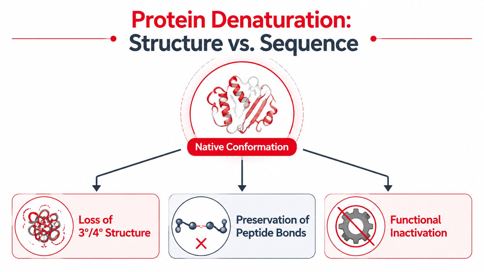
You're reviewing amino acids, enzymes, and protein structure when a question suddenly asks whether a protein has been destroyed, unfolded, or merely separated into subunits. That distinction is the heart of denaturation biochemistry. Protein denaturation is the loss of a protein's native three-dimensional conformation, usually with loss of function, while the peptide backbone generally remains intact.
For medical and premedical exams, the fastest route is to identify the stressor, determine which stabilizing interaction it disrupts, and then ask whether the protein can regain its native fold. Heat, pH changes, chaotropic chemicals, detergents, reducing agents, pressure, and heavy metals can produce related but not identical outcomes.
What Protein Denaturation Actually Means
Denaturation is a conformational change, not automatically chemical destruction. A denatured protein has lost the arrangement required for normal activity, binding, transport, or structural support. In the standard biochemical model, the amino acid sequence remains connected by peptide bonds, but the secondary, tertiary, or quaternary arrangement changes.
That distinction separates denaturation from hydrolysis. Hydrolysis cleaves peptide bonds and changes the primary structure. Denaturation usually disrupts weaker interactions that maintain folding, so the protein can lose function even though its covalent amino acid chain remains present. The historical importance of this distinction dates to 1931, when Hsien Wu proposed that denaturation represented unfolding of a soluble protein rather than hydrolysis or anhydride formation. (Historical account of Wu's theory)
Four structural levels
A useful mental model begins with the four levels of protein structure:
- Primary structure is the linear amino acid sequence joined by peptide bonds.
- Secondary structure includes alpha helices and beta sheets, stabilized mainly by hydrogen bonds involving the peptide backbone.
- Tertiary structure is the complete three-dimensional fold of one polypeptide, supported by hydrophobic interactions, hydrogen bonds, ionic interactions, dispersion forces, and sometimes disulfide bonds.
- Quaternary structure describes the organization of multiple polypeptide subunits.
Denaturation most directly disrupts secondary, tertiary, and quaternary structure. The primary sequence usually survives unless the stress becomes chemically destructive. A protein can therefore retain its full sequence while losing the active-site geometry that made it an enzyme.
For a broader review of the hierarchy, use this guide to primary, secondary, tertiary, and quaternary protein structure. On a board-style question, the key question isn't “Was the protein damaged?” It's “Which level changed, and were peptide bonds cleaved?”
How Denaturation Disrupts Each Structural Level
Protein folding reflects a balance among many weak forces rather than one permanent lock. Hydrogen bonds help organize helices and sheets. Hydrophobic side chains tend to cluster away from water, while ionic interactions, dispersion forces, and disulfide bridges help maintain the mature fold.
The native state is also less thermodynamically distant from unfolding than many students expect. Near physiological temperature, the free-energy gap between native and denatured states is typically about 25–60 kJ·mol⁻¹, while the associated enthalpy and entropy changes are much larger, roughly 500–5000 kJ·mol⁻¹. (Thermodynamic overview of protein stability) This explains why a modest shift in temperature, pH, or denaturant concentration can move a protein across a sharp unfolding threshold.
Cause-to-structural-effect table
| Structural Level | Stabilizing Forces | Effect of Denaturation | Primary Structure Intact? |
|---|---|---|---|
| Primary | Peptide bonds | Usually unchanged during ordinary denaturation | Usually yes |
| Secondary | Backbone hydrogen bonds | Alpha helices and beta sheets lose organization | Yes |
| Tertiary | Hydrophobic interactions, ionic interactions, hydrogen bonds, dispersion forces, disulfide bridges | The single-chain three-dimensional fold is disrupted | Yes, unless chemical cleavage occurs |
| Quaternary | Interactions among separate subunits | Subunits may dissociate or lose their normal arrangement | Each subunit's sequence usually remains intact |
A reducing agent illustrates an important nuance. It can disrupt disulfide bonds, which are covalent links between cysteine residues, without hydrolyzing the peptide backbone. By contrast, a chaotrope weakens the forces that support the hydrophobic core. Reviewing polar amino acids and their interactions can help connect side-chain chemistry to folding behavior.
Exam rule: If the vignette describes loss of activity with preserved peptide bonds, think denaturation. If it describes cleavage of the polypeptide chain, think hydrolysis or proteolysis.
Denaturing Agents and Their Molecular Mechanisms
Different stressors attack different parts of the folding network. That's why “heat breaks proteins” is an incomplete explanation. Heat primarily increases molecular motion, whereas pH changes alter charge, and chemical reagents change solvent behavior or specific covalent interactions.
Heat
Heating increases atomic vibration and can disrupt relatively weak hydrogen bonding and dispersion forces. A standard biochemistry reference identifies temperatures above 50°C as capable of disrupting these interactions. (Protein denaturation reference)
As the fold loosens, hydrophobic groups become more exposed to water. Enzymatic activity may fall over a narrow temperature range because the active site loses its precise geometry before the entire protein appears visibly unfolded. Heating can also promote aggregation, which makes recovery less likely.
pH extremes
Acidic or basic conditions change the ionization state of amino acid side chains. That alters ionic interactions and hydrogen bonding, and similarly charged groups may repel one another. A protein can therefore lose its native fold without any initial break in its peptide bonds.
Chaotropes and detergents
Urea and guanidinium-based reagents weaken the interactions supporting the hydrophobic core. Sodium dodecyl sulfate, or SDS, is an anionic detergent that binds along polypeptide chains, disrupts native hydrophobic interactions, and gives proteins a strong negative charge. SDS-PAGE therefore evaluates proteins after chemical denaturation rather than preserving their native shape.
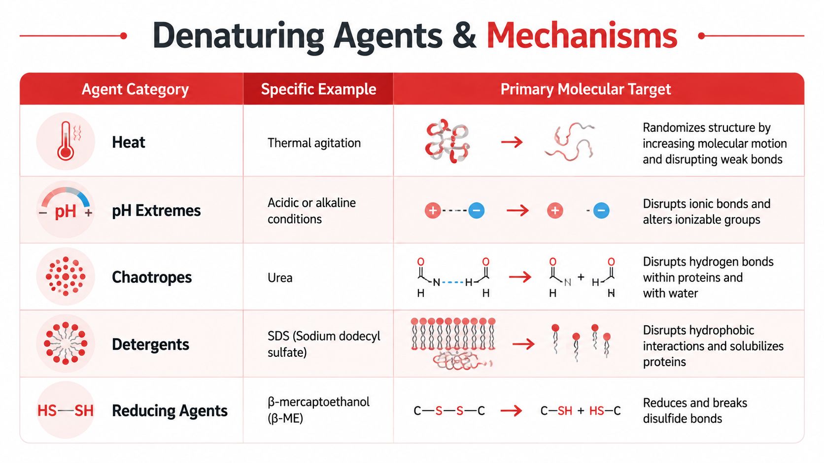
Reducing agents such as beta-mercaptoethanol and dithiothreitol, or DTT, target disulfide bonds. They shouldn't be confused with urea. Urea disrupts noncovalent folding forces, while a reducing agent changes the redox state of cysteine linkages.
Heavy metals and organic solvents
Heavy metal ions, including mercury, silver, and lead, can bind carboxylate groups or cysteine sulfhydryl groups. This can disrupt ionic interactions and disulfide linkages, with effects that may be more persistent than simple reversible unfolding. (Chemical mechanisms of denaturation)
Organic solvents alter the solvent environment around side chains and can disturb hydrophobic packing and electrostatic interactions. The practical exam move is to match the agent with its most specific target rather than assuming every reagent causes the same structural change.
For a medical-student review of the surrounding subject, see this biochemistry guide for medical students.
Reversible Versus Irreversible Denaturation
A protein unfolded during a laboratory procedure may still recover its native conformation after the stressor is removed. This is reversible denaturation, and it is most likely when the chain remains chemically intact, avoids aggregation, and has not formed incorrect covalent bonds.
The change can happen abruptly. Although the free-energy difference between folded and unfolded states may be modest, folding interactions behave cooperatively. A small shift in conditions can disrupt many interactions together, causing a sharp loss of function rather than a gradual decline.
Chemical denaturation is often represented by a near-linear model:
ΔG° = ΔG°w − m[denaturant]
In this equation, ΔG°w is the extrapolated unfolding free energy in water. The m-value describes how strongly unfolding responds to denaturant concentration. A larger m-value generally indicates that unfolding exposes more solvent-accessible surface area, consistent with greater burial of hydrophobic groups in the native state. (Chemical denaturation model)
Why unfolded proteins may not refold
An unfolded chain can encounter another unfolded or misfolded chain and form an aggregate. Heat or chemical stress can also promote side reactions, alter covalent chemistry, or produce incorrect disulfide pairing. Once one of these changes occurs, removing the original stressor may no longer restore the starting structure.
These terms describe related but separate events. Unfolding removes the native three-dimensional conformation. The exposed chains may then associate through aggregation. Chemical modification changes the protein's covalent chemistry, while degradation breaks the polypeptide into smaller fragments. A single sample can show more than one outcome, so loss of activity alone does not identify which process occurred.
Cold and pressure denaturation
Heating is only one route to denaturation. Cold denaturation can result when temperature shifts destabilize hydrophobic hydration and related forces. Pressure can also produce different outcomes under different conditions. Historical work reported egg white coagulation under 7,000 atm within 30 minutes, while later work found coagulation above 3,880 kg/cm². (Historical pressure-denaturation review)
Pressure may also retard thermal denaturation. Mid-20th-century observations described this effect at about 1,000 atm, indicating that pressure can stabilize native structure in some settings. For board-style reasoning, evaluate the protein, solvent, temperature, pressure, and exposure conditions before predicting whether denaturation will be reversible.
Laboratory Methods for Detecting Denaturation
No single assay answers every structural question. A useful method measures a specific consequence of unfolding, such as altered migration, secondary-structure loss, thermal transition, or reduced activity.
SDS-PAGE
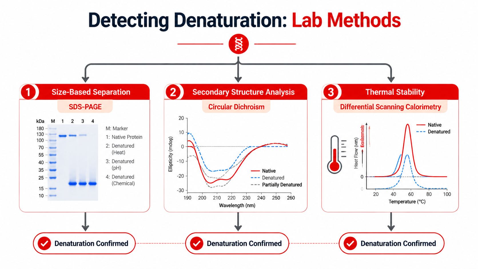
In SDS-polyacrylamide gel electrophoresis, SDS denatures proteins and coats them with negative charge. The gel then separates the treated polypeptides primarily by mass, not by native shape or original charge. A reducing agent may be added when the investigator also wants to disrupt disulfide bonds.
This means SDS-PAGE can confirm that polypeptides differ in apparent molecular mass, but it doesn't directly tell you whether the protein retained its native active conformation.
Circular dichroism
Circular dichroism, or CD, spectroscopy detects changes in the absorption of circularly polarized light. Because alpha helices and beta sheets produce characteristic signals, CD can reveal a shift in secondary structure during unfolding.
CD is especially useful when the question asks whether helices or sheets changed, rather than whether the protein's total mass changed.
Thermal and functional assays
Differential scanning calorimetry, or DSC, measures heat absorbed as a protein passes through a thermal transition. The midpoint temperature, Tm, is the temperature at which native and unfolded populations are equal and ΔG° equals zero. Differential scanning fluorimetry, or DSF, uses a fluorescent signal that changes as hydrophobic regions become exposed during heating.
An enzyme activity assay measures the functional endpoint. If catalytic activity disappears while the protein remains detectable, the result supports a structure-function change rather than complete degradation.
For a focused comparison of SDS-PAGE and gel electrophoresis, keep the central distinction in mind. SDS-PAGE is a denaturing, mass-oriented separation method, while native methods attempt to preserve conformation and activity.
Board-Style Reasoning With a Worked Example
Hypothetical example, not an official or recalled question: A laboratory incubates an enzyme in a strongly acidic solution. The enzyme loses catalytic activity, but analysis shows that its polypeptide chain remains intact. After the solution is returned toward its original pH, activity doesn't fully recover.
Use a fixed sequence instead of chasing the most familiar word in the vignette.
- Identify the stressor. The stressor is an extreme pH, not heat, SDS, or a reducing agent.
- Map the molecular target. The pH shift changes side-chain ionization. Ionic interactions and hydrogen bonds can be disrupted, and like-charge repulsion can destabilize the fold.
- Predict the structural outcome. Secondary and tertiary structure may change, altering the active site. The primary sequence remains intact unless the question provides evidence of peptide-bond cleavage.
- Explain incomplete recovery. The unfolded enzyme may have aggregated or undergone an irreversible change. Denaturation is therefore not automatically reversible.
- Eliminate confounders. Hydrolysis would imply cleavage of peptide bonds. SDS would add detergent-mediated unfolding and negative charge. DTT would specifically reduce disulfide bonds. A chaotrope would weaken hydrophobic-core stabilization.
The same reasoning transfers to a different hypothetical: if a reagent specifically reduces cystine bridges, prioritize disulfide disruption rather than claiming that peptide bonds were hydrolyzed. For a broader approach to critical thinking for medical exams, practice stating the causal chain aloud: stressor, interaction, structural level, function, reversibility.
Reasoning checkpoint: The correct answer usually names both the affected interaction and the preserved feature. “The protein unfolded” is less precise than “pH altered side-chain ionization, disrupting tertiary interactions while leaving the peptide backbone intact.”
Clinical Relevance and High-Yield Study Framework
A protein can lose activity during a heat exposure even while remaining visibly clear. The active site may shift before precipitation appears, so clinical and laboratory interpretation must separate functional loss from gross aggregation. Heat can inactivate enzymes and alter food proteins. Disinfectants and sterilization procedures disrupt protein structure, while heavy metals can bind sulfur-containing or carboxylate-containing groups.
Therapeutic proteins add a practical distinction: temporary unfolding may permit recovery after the stressor is removed, whereas aggregation or chemical modification can prevent refolding. Reviews of therapeutic protein stability therefore examine recovery after heat or urea exposure rather than assuming every denaturation event is permanent. (Therapeutic protein denaturation overview)
Use this synthesis checklist for a board-style question or laboratory result:
- Separate activity from appearance. Loss of catalytic function can precede visible precipitation because active-site geometry is more sensitive than overall solubility.
- Set the thermodynamic threshold. Ask whether the stress stayed below the point where native interactions can reform. Removal of the agent may allow refolding, but crossing a threshold that promotes aggregation or chemical change can make recovery incomplete.
- Match the test to the outcome. Spectroscopy can reveal altered secondary structure, thermal analysis can show changed stability, solubility assays can detect aggregation, and an activity assay tests functional recovery.
- State the preserved feature. A strong answer identifies what changed and what remains sufficiently intact, then explains why function did or did not return.
Frequently asked questions
How does denaturation differ from degradation?
Denaturation involves loss of native conformation while peptide bonds remain intact. Degradation involves chemical breakdown, including possible cleavage of the polypeptide chain.
Can chaperone proteins reverse denaturation?
Chaperones can assist folding and reduce inappropriate aggregation in biological systems. Recovery still depends on whether the protein retains a chemically intact sequence and can cross back to its native state.
Why can a protein lose function before it visibly precipitates?
Catalysis requires precise active-site geometry. A small conformational shift can disrupt substrate binding or catalytic positioning before enough unfolded protein accumulates to scatter light.
Is misfolding always classical denaturation?
Misfolding can arise from altered folding pathways, mutations, cellular stress, or impaired quality control. It overlaps with denaturation but covers a broader range of structural problems.
Ace Med Boards offers diagnostic tutoring and personalized study-plan support for learners connecting biochemistry mechanisms with board-style reasoning. Visit Ace Med Boards to review its focused consultation option.