You're staring at a labeled anatomy diagram, recognizing individual names but struggling to explain what would happen if one structure were injured. That gap is exactly what board-style questions exploit. Anatomy becomes more manageable when you organize it around spatial relationships, function, and clinical consequences instead of memorizing isolated labels.
This guide builds that framework for medical students, osteopathic medical students, international medical graduates, residents, and learners preparing for foundational examinations. It begins with body systems, directional language, and planes, then moves to surface landmarks, common traps, study methods, and original clinical reasoning examples.
Why Anatomy Still Matters for Your Board Scores
Anatomy becomes more manageable when you organize it around spatial relationships, function, and clinical consequences. That approach turns a nerve, vessel, or compartment into a clinical coordinate system rather than a disconnected label. It also mirrors board questions, which may present a structure directly or ask for the deficit caused by trauma several centimeters away.
A nerve has a route, motor function, sensory territory, and vulnerable relationships. A vessel has branches, supplied tissues, anastomoses, and consequences when flow is interrupted. A compartment has boundaries, contents, and pressure-related risks. Linking these features makes recall more durable because each fact has a reason to exist.
Practical rule: For every structure, ask where it is, what it does, what travels with it, and what finding follows when it is damaged.
The adult skeleton is commonly taught as 206 bones, although the count can vary because some people have extra sesamoid bones, accessory ossicles, an extra rib, or an extra vertebra, as described by Britannica's overview of the human body. Anatomy's modern observation-based tradition also owes a historical debt to Andreas Vesalius. His 1543 publication, De Humani Corporis Fabrica, relied largely on human dissection and challenged dependence on inherited classical texts, according to the same reference.
Build an exam-oriented mental map
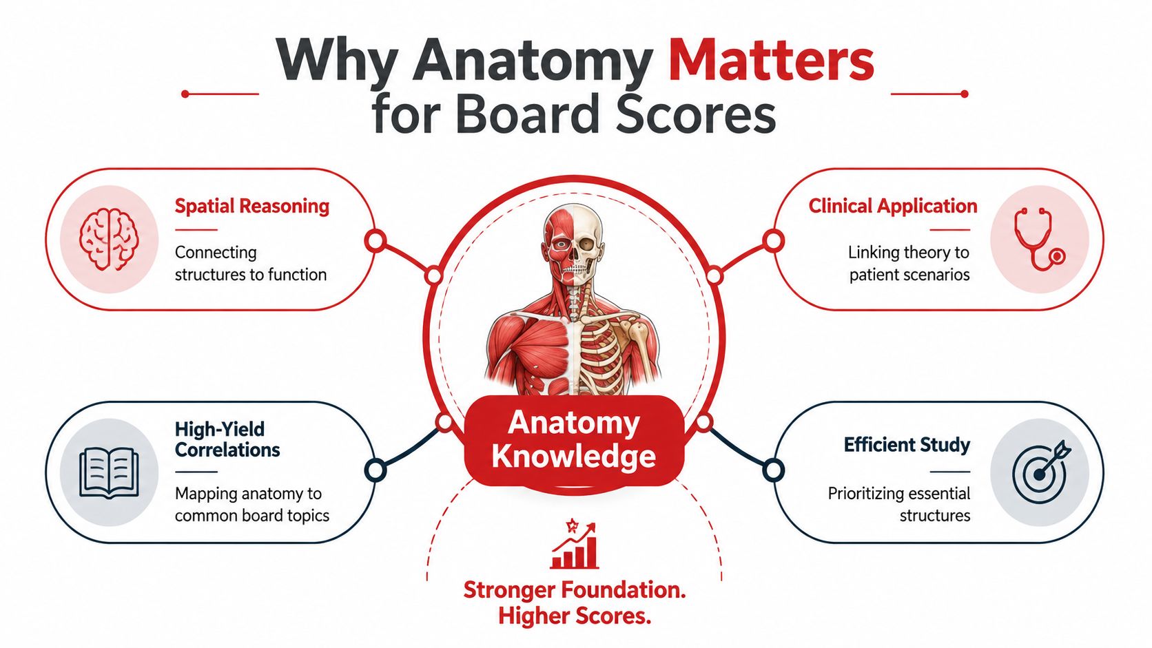
A strong anatomy review connects four layers:
- Spatial reasoning: Locate structures relative to planes, compartments, landmarks, and neighboring tissues.
- Function: Link structure to movement, conduction, drainage, ventilation, filtration, or protection.
- Clinical application: Predict deficits, imaging findings, procedural risks, and patterns of referred symptoms.
- Question strategy: Identify the lesion location before choosing the diagnosis or named structure.
The USMLE content outline helps place gross anatomy and embryology within a broader study plan. Use it to connect regional review with the clinical reasoning expected on board examinations.
Foundational Anatomical Framework and Directional Language
A patient may lie on an examination table, rotate during imaging, or present with a flexed limb. Anatomical descriptions still begin from one fixed reference, the standard anatomical position. The person stands erect with the hips and knees extended, head facing forward, eyes open and looking straight ahead, and mouth closed, as explained by Radiopaedia's anatomical-position reference.
This shared reference keeps clinical language consistent. A flexed arm still has an elbow distal to the shoulder. A lesion may appear on the left side of an image, yet its description depends on patient orientation and the conventions used for that study. The framework functions like a map legend: it allows every reader to interpret the same relationship.
Learn the language in pairs
| Term | Definition | Clinical Example |
|---|---|---|
| Superior, or cranial | Toward the head | The thorax is superior to the abdomen. |
| Inferior, or caudal | Away from the head | The pelvis is inferior to the abdomen. |
| Anterior, or ventral | Toward the front | The sternum is anterior to the heart. |
| Posterior, or dorsal | Toward the back | The vertebral column is posterior to the heart. |
| Medial | Toward the body midline | The ulna is medial to the radius in anatomical position. |
| Lateral | Away from the body midline | The lungs are lateral to the mediastinum. |
| Proximal | Nearer the trunk or point of origin | The elbow is proximal to the wrist. |
| Distal | Farther from the trunk or point of origin | The fingers are distal to the elbow. |
These definitions follow the National Cancer Institute's anatomy terminology guide. Use precise directional terms rather than general words like “above,” “below,” or “near.” On an exam, that precision helps you translate a stem into a structural relationship before selecting a diagnosis.
Treat planes as your imaging coordinate system
The sagittal plane separates left from right. A midsagittal section passes through the midline, while a parasagittal section lies beside it. The coronal plane separates anterior from posterior. The transverse, or axial, plane separates superior from inferior.
A frequent trap is confusing the plane with the direction of a structure. A transverse image is not itself “inferior” or “superior.” Locate the structure within the slice, then trace it across adjacent slices. Pair each plane with a task: sagittal images clarify midline relationships, coronal images support front-to-back comparison, and axial images help localize structures in cross-section.
For a focused review, use Anatomical Terms and Directions, then describe structures from memory without viewing a labeled diagram. This converts terminology from passive recall into a spatial reasoning skill.
Organizing the Body Into High-Yield Systems
A patient with shortness of breath may test anatomy across several systems at once. To reason through the case, connect the airway and lungs with thoracic mechanics, blood flow, autonomic input, and acid-base effects. Anatomists commonly describe the body through 11 organ systems. Some combine the skeletal and muscular systems as one musculoskeletal system and count 10, according to Human Body Parts Anatomy. Regardless of how many systems you count, the relationships between them matter more than the classification itself.
The cardiovascular system supplies tissues, the respiratory system exchanges gases, and the renal system regulates fluid and electrolyte balance. The nervous and endocrine systems coordinate responses. The integumentary and lymphatic systems provide barriers, drainage, and immune surveillance. The digestive and reproductive systems rely on vascular, neural, muscular, and connective-tissue support.
Use a system map, not a list
| System | Structural focus | Function to anchor |
|---|---|---|
| Nervous | Brain, spinal cord, cranial and peripheral nerves | Communication and control |
| Endocrine | Hormone-producing glands and target tissues | Chemical regulation |
| Cardiovascular | Heart, arteries, veins, and capillaries | Transport and perfusion |
| Respiratory | Airways, lungs, and thoracic mechanics | Ventilation and gas exchange |
| Digestive | Gastrointestinal tract and accessory organs | Processing and absorption |
| Urinary | Kidneys, ureters, bladder, and urethra | Filtration and homeostasis |
| Reproductive | Gonads, ducts, supporting structures | Reproduction and hormonal function |
| Musculoskeletal | Bones, muscles, joints, and connective tissues | Movement, stability, and protection |
| Integumentary | Skin and associated structures | Barrier and sensation |
| Lymphatic and immune | Lymphatic vessels, nodes, spleen, and immune tissues | Drainage and defense |
| Sensory structures | Specialized visual, auditory, and other receptors | Environmental input |
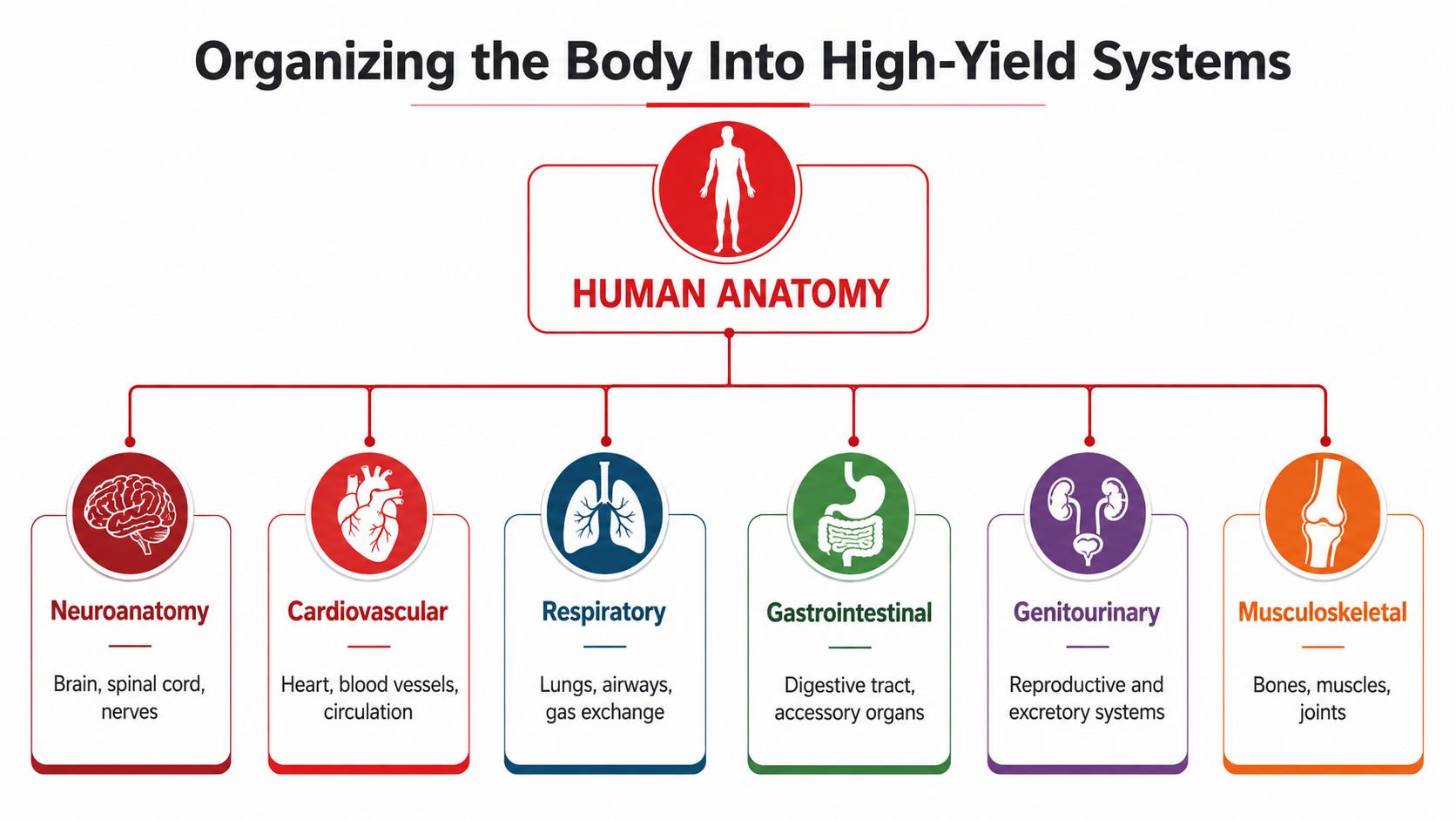
The body also contains more than 600 muscles, and some anatomy references place the musculoskeletal system at roughly 35% to 45% of body weight, as summarized by the same anatomy reference. These figures explain why movement, trauma, compartment relationships, and structural pathology appear repeatedly in medical education. Use the diagram of six major high-yield organ systems to rehearse how regions connect rather than memorizing each label separately.
Follow the function across systems
A renal question may depend on retroperitoneal relationships, arterial branching, embryologic ascent, and endocrine regulation. Build each system around a four-part chain:
- Structure: What is present?
- Route: Where does it travel or drain?
- Function: What does the route permit?
- Failure pattern: What changes when the structure is interrupted?
That chain turns a collection of labels into a clinical reasoning scaffold. It also exposes exam traps, because a lesion's location, pathway, and lost function must agree.
High-Yield Systems and Their Clinical Correlations
High-yield anatomy is not a ranking of isolated structures. It's the study of relationships that let you infer a deficit from a lesion, an imaging finding from a plane, or a procedural risk from a landmark.
Neuroanatomy
Start with pathways and localization. For a peripheral nerve, connect root levels to plexus organization, terminal nerve, muscles supplied, and sensory distribution. For a central lesion, identify whether the pattern points to cortex, brainstem, spinal cord, or a long tract. The question often gives you the deficit first and expects you to reason backward.
A useful example is a nerve injury near a joint. Don't memorize only the nerve's name. Ask which muscles lose function, what movement becomes weak, whether sensation changes, and which neighboring structure could explain an alternative answer.
Cardiovascular and respiratory anatomy
Cardiovascular questions reward spatial tracing. Follow blood from chamber to valve to vessel, then connect a territory to its supplied tissue. Coronary anatomy becomes more memorable when you pair each arterial course with the myocardial region and electrical or mechanical consequence of ischemia.
Respiratory anatomy depends on branching and compartments. Know how the trachea divides, how bronchi relate to lung structures, and how bronchopulmonary segments support localized disease or surgical planning. The same reasoning applies to pleural spaces, mediastinal relationships, and the neurovascular structures of the thoracic wall.
For a related physiology connection, review cardiovascular physiology concepts while tracing the structures that make those functions possible.
Gastrointestinal, genitourinary, and musculoskeletal anatomy
In the abdomen, use layers and compartments. Identify peritoneal relationships, vascular territories, lymphatic drainage, and the route of a structure through the abdominal wall or retroperitoneum. For the genitourinary system, organize the kidneys, ureters, bladder, reproductive organs, and pelvic floor around flow, support, and shared spaces.
Musculoskeletal questions become easier when you connect origin, insertion, action, and innervation. Then add the joint position that increases or decreases tension and the nearby nerve or vessel vulnerable to trauma. Head and neck anatomy follows the same principle, but with tighter spaces and more overlapping cranial nerve, vascular, airway, and fascial relationships.
Reasoning pattern: Localize first, name second. A correct structure without a defensible location is fragile recall.
Surface Landmarks and Common Board Question Traps
Surface anatomy connects diagrams to examination, imaging, regional anesthesia, and procedural planning. But traditional landmarks aren't equally reliable in every person. An evidence-based reappraisal found substantial variation in the course of the spinal accessory nerve, including its exit from the sternocleidomastoid and entry into the trapezius, as reported in the University of Otago research record.
High-yield surface landmarks
| Region | Landmark | Clinical Significance |
|---|---|---|
| Antecubital | Anterior elbow region | Helps localize structures used in examination and venous access. |
| Popliteal | Posterior knee region | Frames the neurovascular structures behind the knee. |
| Inguinal | Groin region | Helps distinguish groin relationships and hernia locations. |
| Umbilical | Navel region | Provides a central abdominal surface reference. |
| Abdominal | Area inferior to the chest and superior to the pelvis | Supports regional localization of abdominal findings. |
| Posterior triangle of the neck | Region bordered by neck and shoulder landmarks | Important for locating structures including the spinal accessory nerve. |
Regional terms such as antecubital, popliteal, inguinal, and umbilical identify body sites directly, as outlined by LibreTexts' surface and regional anatomy reference.
Traps that turn knowledge into wrong answers
Trap one, treating a textbook landmark as an absolute coordinate. The spinal accessory nerve's relationship to neck landmarks varies widely. For real clinical decisions, context, palpation, imaging, and direct confirmation matter. Board questions may test the principle that a classic landmark isn't infallible.
Trap two, reversing proximal and distal. These terms refer to the trunk or origin, not the direction the limb happens to point. Establish the reference structure before interpreting the wording.
Trap three, ignoring variants. A systematic review recommends greater emphasis on anatomical variation in medical education, while a later scoping review found that only 25.5% of reported gross-anatomy teaching approaches explicitly addressed variation, according to the PubMed-indexed review. When a vignette seems inconsistent with the standard model, ask whether the variant changes the diagnosis or procedure.
Trap four, reading slices as labels. Cross-sectional anatomy requires orientation, continuity, and three-dimensional reconstruction. Don't identify a circular structure from one image alone. Trace it through neighboring slices and verify its relationships.
Evidence-Based Study Strategies for Anatomy Mastery
Passive rereading feels productive because the page looks familiar. Recognition, however, isn't the same as retrieval. A stronger session asks you to produce the pathway, label a blank diagram, explain a deficit, or identify a structure in an axial image before checking the answer.
Cross-sectional anatomy deserves special attention. In one survey, 74% of resident physicians reported no formal cross-sectional anatomy training, while 88% reported high clinical needs; positioning was the top challenge for 45% of respondents, according to the study in PubMed Central. The same report noted that fewer than 40% of medical schools worldwide provide systematic cross-sectional anatomy training to all students.
A practical weekly framework
Adapt the sequence to your examination and schedule:
- Map: Begin with one region or system. Draw compartments, routes, and major relationships from memory.
- Retrieve: Use blank diagrams, flashcards, or oral prompts without notes.
- Integrate: Complete original or licensed practice questions, then classify each error as location, function, terminology, variant, or reasoning.
- Revisit: Schedule spaced reviews so the same material returns at increasing intervals. Spaced repetition for medical study can help you build that cycle.
- Teach: Explain one pathway aloud in plain language, including what changes after injury.
Short, focused learning blocks can also support repeated retrieval. If you're comparing digital formats, a neutral overview of microlearning platforms for corporate training may help you evaluate features such as brief lessons, review prompts, and progress organization, while remembering that medical accuracy still depends on the content source.
Readiness checklist
Before moving on, confirm that you can:
- Orient: Identify anatomical position, plane, and patient side.
- Localize: Predict the structure involved from a motor, sensory, vascular, or imaging finding.
- Connect: Explain how structure supports function.
- Handle variation: Recognize when a standard landmark or pathway may differ.
- Review errors: State why each distractor is wrong, not only why the answer is right.
Inclusive anatomy also matters. A 24-country campaign reported that 1 in 3 students and 1 in 4 staff didn't feel represented in anatomy programs, while recent education research describes a continued default toward a narrow body type, according to Frontiers in Education. Study resources should support anatomical variation, clear language, and clinically relevant representation.
Sample Practice Questions and Reasoning Walkthroughs
The following examples are composite educational illustrations, not official, recalled, or copied examination questions. Their purpose is to model the reasoning sequence.
Question one
A patient develops shoulder drooping and difficulty shrugging after a posterior-triangle neck procedure. Which nerve is most likely injured?
Reasoning: Shoulder shrugging depends on trapezius function. Trapezius is supplied by the spinal accessory nerve. The nerve crosses the posterior triangle and has variable surface relationships, so the deficit localizes the injury more reliably than a memorized skin landmark. The answer is the spinal accessory nerve.
Question two
An imaging vignette describes a lesion in the posterior aspect of the thorax, behind the heart. Which directional term best describes the lesion's relationship to the heart?
Reasoning: “Behind” corresponds to posterior, or dorsal. Don't substitute inferior just because the lesion appears lower on a particular image. Establish the anatomical reference first, then interpret the plane and location.
Question three
A patient has weakness of a movement performed by a muscle, but sensation remains intact. How should you approach the question?
Reasoning: Separate motor from sensory function. Identify the muscle's action, determine its innervation, and localize the lesion along the motor pathway. Then compare nearby nerves and roots that could produce a similar deficit. This method prevents choosing a nerve based only on regional proximity.
For additional practice with this format, use the original examples in Ace Med Boards' Step 1 sample question resource. Practice questions should supplement, not replace, official examination guidance, approved learning materials, and your own error review.
Medical anatomy content here is for examination education, not diagnosis or individualized medical advice. Clinical decisions require patient-specific assessment and current professional guidance.
Ace Med Boards offers diagnostic tutoring and personalized study-plan support for learners who need to connect anatomy with board-style reasoning, including USMLE and COMLEX preparation. Visit Ace Med Boards to explore the available support and request a low-pressure consultation about your current anatomy gaps.