206 bones and roughly 37.2 trillion cells provide useful anchors for anatomy and physiology, but board preparation depends on connecting structure to function and then predicting the lesion. Anatomy identifies the body's structures, while physiology explains what those structures do, so exam questions test them together through clinical reasoning rather than as separate memorization lists.
You may know the name of a nerve, recognize a pressure-volume loop, and still miss a question because you didn't connect the two. A lesion's location changes which fibers are affected, and that anatomical detail determines the physiological deficit. The same reasoning applies whether you're studying a cranial nerve, nephron segment, cardiac conduction pathway, or respiratory control center.
Why Anatomy and Physiology Are One Subject on Boards
Anatomy is the study of structure. Physiology is the study of how those structures work, including the chemistry and physics that support life. Structures can be large enough to see without magnification or small enough to require a microscope, which is why anatomy overlaps with histology and cellular physiology (OpenStax explains the relationship between anatomy and physiology).
Consider a patient with weakness of the tongue after a vascular event. If you identify only the affected cranial nerve but ignore the neighboring motor pathway and the artery supplying the region, your physiological conclusion may be wrong. The board-style task isn't merely naming a structure. It's deciding how location changes function.
The reasoning loop
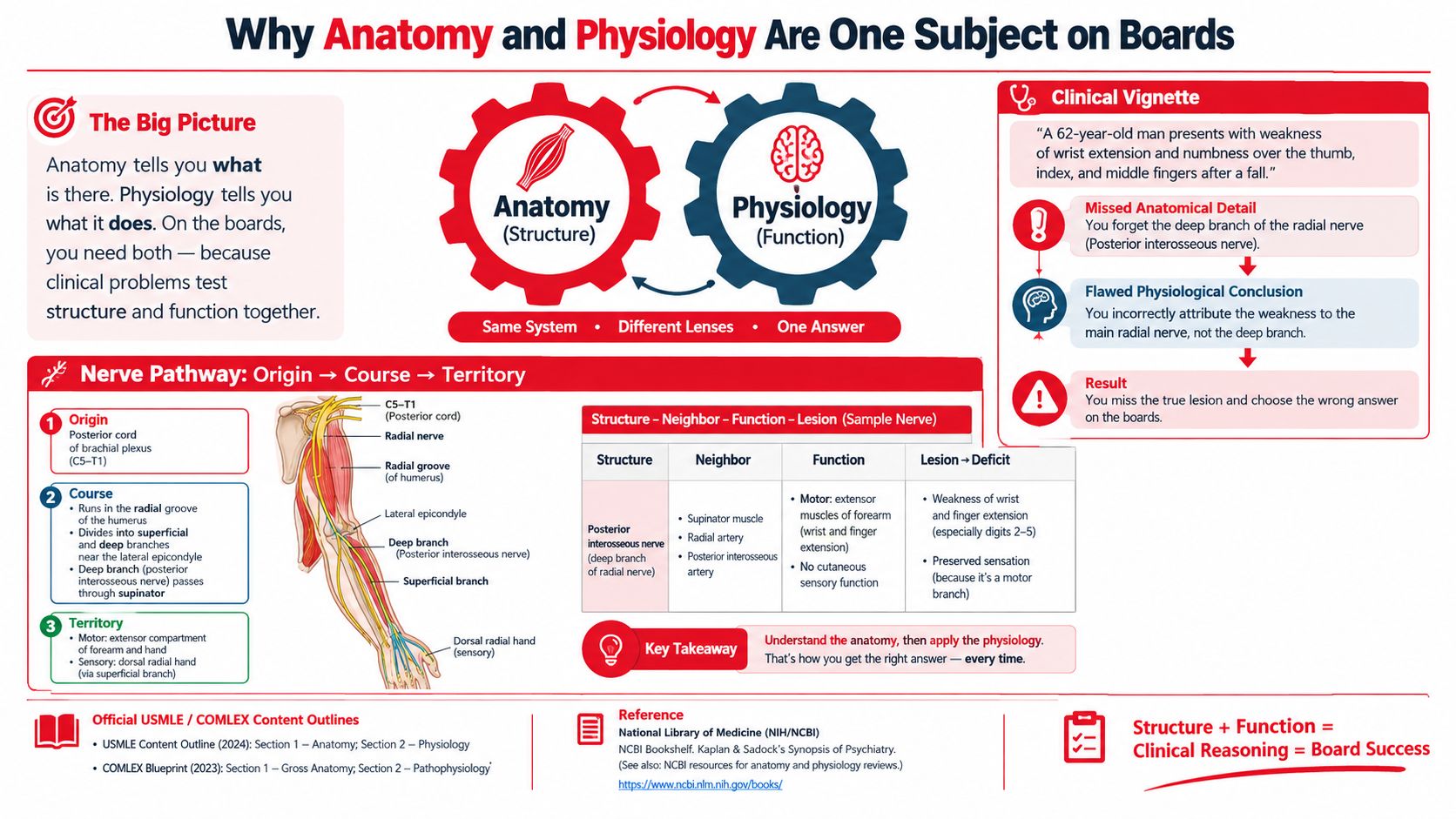
Use four questions whenever a stem presents a lesion, laboratory value, or physiological change:
- Structure: What exact structure is involved?
- Neighbor: What lies beside it, travels with it, or shares its blood supply?
- Function: What does the structure normally do?
- Lesion: What deficit should appear if that structure or its neighbor is damaged?
Anatomy supplies the map: location, boundaries, relationships, blood supply, and innervation. Physiology supplies the operating rules: gradients, feedback loops, resistance, compliance, filtration, secretion, and rate-limiting steps.
That integration is central to the way the United States Medical Licensing Examination (USMLE) tests basic science. The official USMLE Step 1 content outline includes both anatomy and physiology among its disciplines. Physiology represents 30% to 40% of Step 1 items, according to the Step 1 content area report, so students can't treat it as a supporting topic.
Practical rule: Don't ask only, “What is this structure?” Ask, “What does its position make possible, and what would its injury remove?”
A focused clinical reasoning framework can help you turn both subjects into one diagnostic habit. You aren't building two unrelated lists. You're learning to move from a three-dimensional map to a functional prediction.
High-Yield System Map Across the Eight Exam Domains
A useful system map begins with the same sequence every time: structure, physiological role, and lesion pattern. This prevents the common mistake of learning a system's anatomy in one chapter and its physiology in another without reconnecting them.
| System | Key Structure | Core Physiology | High-Yield Lesion |
|---|---|---|---|
| Cardiovascular | Atrioventricular (AV) node | Delays conduction so the ventricles can fill before contraction | AV conduction block |
| Respiratory | Carotid bodies | Detect arterial oxygen changes and contribute to ventilatory regulation | Impaired peripheral chemoreceptor response |
| Nervous | Cranial nerve nuclei and long tracts | Route motor, sensory, and autonomic information | Crossed brainstem deficits |
| Gastrointestinal | Sphincter of Oddi | Regulates delivery of bile and pancreatic secretions into the intestine | Obstructed outflow |
| Renal | Loop of Henle | Establishes a medullary osmotic gradient through segment-specific transport | Impaired urine concentration |
| Endocrine | Adrenal cortex zones | Produces steroid hormones through region-specific enzyme expression | Zone-specific hormone deficiency |
| Musculoskeletal | Rotator cuff | Stabilizes the humeral head during shoulder movement | Weakness or instability from tendon injury |
| Reproductive | Seminiferous tubules | Support spermatogenesis within the testicular environment | Reduced sperm production |
Cardiovascular and respiratory anchors
In the cardiovascular system, pair conduction anatomy with pressure and flow. The AV node is not just a labeled point on a diagram. Its position and conduction properties explain the delay between atrial and ventricular activation. The same approach applies to pressure-volume loops, where chamber structure and valve position determine how pressure and volume change through the cardiac cycle.
For respiratory questions, connect lung regions to ventilation-perfusion relationships and connect chemoreceptor location to the stimulus each receptor detects. A student who remembers only a diagram may confuse oxygen sensing with carbon dioxide and pH regulation. A student who identifies the receptor, its location, and its physiological input is more likely to choose the correct mechanism.
The remaining systems
Neuroanatomy requires attention to tracts, nuclei, and neighbors. Gastrointestinal questions become clearer when you pair each secretion or movement with its anatomical site. Renal questions depend on matching each nephron segment with its transport function, especially the loop of Henle.
Endocrine reasoning improves when gland zones, hormone synthesis, and feedback are kept together. In musculoskeletal anatomy, a joint's stabilizers explain its movement limits and injury patterns. Reproductive physiology similarly depends on linking gonadal anatomy, seminiferous tubules, and hormonal timing. For a focused review of hormonal pathways, use this endocrine system study guide.
The Misconceptions That Cost Points and Why They Persist
Cardiovascular and respiratory misconceptions often survive formal instruction. A review describes misconceptions as persistent and resistant to change, including misunderstandings of the heart-lung circulation relationship, breathing mechanics, and teleological explanations of respiration (review of persistent misconceptions in medical education).
One recurring error is treating oxygen as the primary driver of normal ventilation in every context. The more reliable approach is to identify the receptor and its stimulus. Central chemoreceptors lie near the ventrolateral medulla and respond mainly to changes in cerebrospinal fluid pH related to carbon dioxide, while peripheral chemoreceptors provide a different sensory route.
Another error involves capillary fluid movement. Students may remember “Starling forces” without separating capillary hydrostatic pressure from plasma oncotic pressure. The result is a diagram-based answer that doesn't explain why fluid leaves or returns to a compartment.
Why memorization doesn't repair the problem
Traditional study methods often isolate the facts. A lecture labels a receptor, a flashcard states its stimulus, and a separate diagram shows a circulation pathway. Each item may be individually correct, but the learner still lacks the relationship that makes the answer predictable.
Research on medical curricula also reports that misconceptions are concentrated in physiology, particularly cardiovascular and respiratory topics (curriculum study of physiology misconceptions). That pattern suggests that adding more isolated facts won't solve the problem. Students need to test whether their mental model predicts the finding.
If your explanation uses “because the body wants to” without naming a sensor, signal, pathway, and target organ, the mechanism probably needs another pass.
The structure-neighbor-function-lesion loop exposes faulty models. Spatial relationships constrain possible answers. A receptor's location limits what it can sense, a vessel's territory limits the deficit from occlusion, and a nephron segment's transporters limit the expected urine change.
The Structure Neighbor Function Lesion Method
Use an original vignette about a medial medullary stroke. A patient develops weakness of the opposite side of the body, loss of position and vibration sense on that side, and weakness with tongue movement toward the side of the lesion. The question isn't asking you to recall a memorized cluster. It is asking you to localize the structures that travel through the medial medulla.
Four deliberate passes
Structure: Identify the corticospinal tract, medial lemniscus, and hypoglossal nucleus or exiting fibers. These structures account for the motor, proprioceptive, and tongue findings.
Neighbor: Place each structure in relation to the others. The corticospinal tract carries descending motor fibers, the medial lemniscus carries ascending discriminative sensation, and hypoglossal fibers serve tongue movement. Their proximity makes a single medial medullary lesion plausible.
Function: Translate each structure into a job. Corticospinal injury produces upper motor neuron weakness below the lesion. Medial lemniscus injury disrupts vibration and proprioception. Hypoglossal involvement produces an ipsilateral tongue deficit.
Lesion: Predict the complete pattern before looking at the answer choices. The lesion should produce findings that match the affected tracts and the side of the cranial nerve deficit.
| Step | Sample Vignette, Medial Medullary Stroke | Template for Any Anatomy Block |
|---|---|---|
| Structure | Identify corticospinal tract, medial lemniscus, and hypoglossal fibers | Name the smallest exact structure that explains the finding |
| Neighbor | Note that these pathways occupy the medial medulla | List adjacent tracts, nuclei, vessels, muscles, or compartments |
| Function | Link each pathway to motor control, proprioception, or tongue movement | State the normal function in one sentence |
| Lesion | Predict contralateral body weakness and sensory loss with an ipsilateral tongue deficit | Predict the deficit before reading the options |
A portable note format
Copy the four labels into your question review:
- Structure: What was tested?
- Neighbor: What nearby structure could create a similar presentation?
- Function: What normal process depends on this location?
- Lesion: What pattern should damage produce?
The same format works for cranial nerves, brachial plexus branches, vascular territories, and joint anatomy. It also supports the broader human anatomy study guide approach of connecting location, function, and clinical application.
A labeled composite learner may memorize a nerve name but miss the lesion because the nerve's origin, course, sensory territory, and neighboring landmarks were never connected. Trace the pathway from origin to target, move the hypothetical injury site, and then ask how the deficit changes. That shift from naming to localization is the point of the method.
Applying the Loop to Physiology Reasoning
Physiology questions often change one variable and ask you to predict the result. Before calculating or choosing an answer, define the mechanism, compare it with its closest look-alike, apply it to the stem, and explain why the distractors fail.
Consider an original hypothetical vignette involving acute blood loss. Reduced circulating volume lowers venous return, which reduces ventricular filling and stroke volume. Baroreceptor-mediated responses then increase sympathetic activity, raising heart rate and contractility while constricting selected vascular beds.
A mechanism-first sequence
- Define the core concept: Less effective circulating volume reduces preload.
- Build a mechanism map: Reduced preload lowers stroke volume, which affects cardiac output unless compensation occurs.
- Compare close look-alikes: Distinguish low preload from high afterload, impaired contractility, or distributive loss of vascular tone.
- Apply one original vignette: Predict the immediate cardiovascular response to the changed variable.
- Explain every distractor: A distractor may describe a response associated with a different primary defect.
- Test transfer: Change one variable, such as contractility rather than volume, and rebuild the prediction.
This sequence also transfers to renal and endocrine questions. For a loop diuretic, identify the nephron segment, its transporter, the affected gradient, and the downstream effect on urine composition. For an endocrine disorder, identify the gland or zone, the hormone produced, the feedback signal, and the expected change in the regulatory axis.
Use these prompts before answering:
- What variable changed first?
- Which structure senses that change?
- What pathway carries the signal?
- What function changes immediately?
- Which neighboring mechanism could produce a similar laboratory pattern?
- What single change would reverse or modify the prediction?
The physiology study guide can serve as a focused companion, but active explanation matters more than rereading. If you can't describe the mechanism without looking at the diagram, the concept isn't ready for timed application.
A Six-Week Study Framework With Error-Log Practice
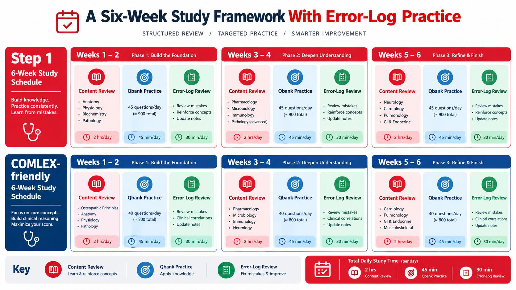
A six-week plan should separate learning, application, and correction without treating them as unrelated tasks. The schedule below is a framework for USMLE Step 1 preparation. Students preparing for COMLEX-USA should preserve the same anatomy and physiology loop while adding the osteopathic content and exam-specific resources required by their plan.
| Phase | Step 1 emphasis | COMLEX-friendly adaptation | Error-log focus |
|---|---|---|---|
| Weeks 1 to 2 | Review foundational anatomy and physiology by system, then complete timed case-based questions | Pair the same systems with osteopathic principles and the resources required for COMLEX-USA | Mislocalized structures and incorrect mechanisms |
| Weeks 3 to 4 | Rotate cardiovascular, respiratory, renal, endocrine, and neuro integration | Keep system integration while adding exam-specific osteopathic review | Repeated distractor patterns and weak links |
| Weeks 5 to 6 | Shift toward mixed blocks, timed application, and targeted repair | Use mixed practice with continued osteopathic integration | Retest errors without relying on recognition |
A practical daily pattern might include two hours of focused review, a timed question block, and a short error-log session. The exact duration should reflect your available study time and current readiness, not an invented universal target.
The error-log workflow
Record every missed or guessed question in these columns:
- System and structure: Name the anatomical region or physiological pathway.
- Tested function: State what the structure normally does.
- Your prediction: Write the answer you chose and why.
- Failure type: Label the error as recall, localization, mechanism, calculation, or premature closure.
- Correct mechanism: Explain the answer in your own words.
- Discriminator: Identify the detail that separates the correct answer from the closest distractor.
- Transfer change: Alter one variable and predict the new result.
- Retest date: Return to the concept without copying the original question.
Error-log standard: A correction isn't complete until you can explain why the correct answer works and why the nearest distractor doesn't.
Use spaced repetition for the final retrieval cue, not as a substitute for mechanism mapping. Interactive medical education can also reinforce three-dimensional relationships, and Studio Liddell's overview of XR medical training offers context for how immersive environments may support spatial learning.
Weekly self-audit
At the end of each week, ask:
- Coverage: Which system still produces repeated errors?
- Localization: Did I identify structures and neighbors before choosing?
- Mechanism: Can I explain the physiological change without a diagram?
- Distractors: Do my misses cluster around one look-alike?
- Transfer: Can I predict the result after changing one variable?
- Timing: Can I perform the reasoning under the official exam's timed conditions?
The USMLE Step 1 format is highly structured, with an 8-hour exam divided into seven blocks, and each block allows up to 40 questions with one hour for the block (exam format overview). Practice should therefore include both untimed learning and timed reasoning.
Key Takeaways and Next Step for Your Prep
- Use one reasoning loop: Move from structure to neighbor to function to lesion whenever a question presents a clinical finding.
- Map systems, don't isolate chapters: Pair the AV node with conduction physiology, the carotid bodies with respiratory control, the loop of Henle with transport, and the adrenal zones with hormonal feedback.
- Repair misconceptions directly: Cardiovascular and respiratory errors deserve dedicated error-log entries because memorized diagrams may preserve faulty mental models.
- Practice transfer: After solving an original vignette, change one variable and predict how the answer changes.
- Review actively: Case-based questions and a specific error log reveal whether you can apply a concept rather than recognize a familiar phrase.
This framework applies to anatomy and physiology preparation for USMLE Step 1 and can be adapted to other board timelines without blending their distinct blueprints or requirements. It also gives you a concrete starting point tonight: choose one missed question, complete the four-column reasoning loop, and retest the mechanism after changing one variable.
Anatomy and physiology study is clinically relevant, but this article is for examination education, not individualized medical advice or patient treatment decisions.
If you want help turning your anatomy and physiology errors into a focused board plan, Ace Med Boards offers personalized online tutoring and exam-preparation support, including case-based review and question-analysis guidance. You can schedule a free consultation to discuss your current needs without committing to a particular study path.