
Hematology becomes manageable when you integrate cell size, marrow response, smear morphology, and the confirmatory mechanism instead of memorizing isolated diagnoses. For anemia, start with the mean corpuscular volume, then ask whether the reticulocyte response is appropriate, inspect the smear, and choose the test that confirms the mechanism.
A tired student can often recognize that a complete blood count is abnormal but still lose the question by reading one value in isolation. A low hemoglobin may reflect impaired production, blood loss, hemolysis, inflammation, or a mixed process. The same disciplined sequence also helps with thrombocytopenia, coagulation abnormalities, leukemia, transfusion reactions, and anticoagulant reversal.
Hematology carries major clinical and examination weight. In 2022, hematologic malignancies accounted for 1,311,104 new cases and 700,205 deaths worldwide (global hematologic malignancy burden). You don't need to memorize every disease at once. You need a reliable way to turn a stem into a sequence of decisions.
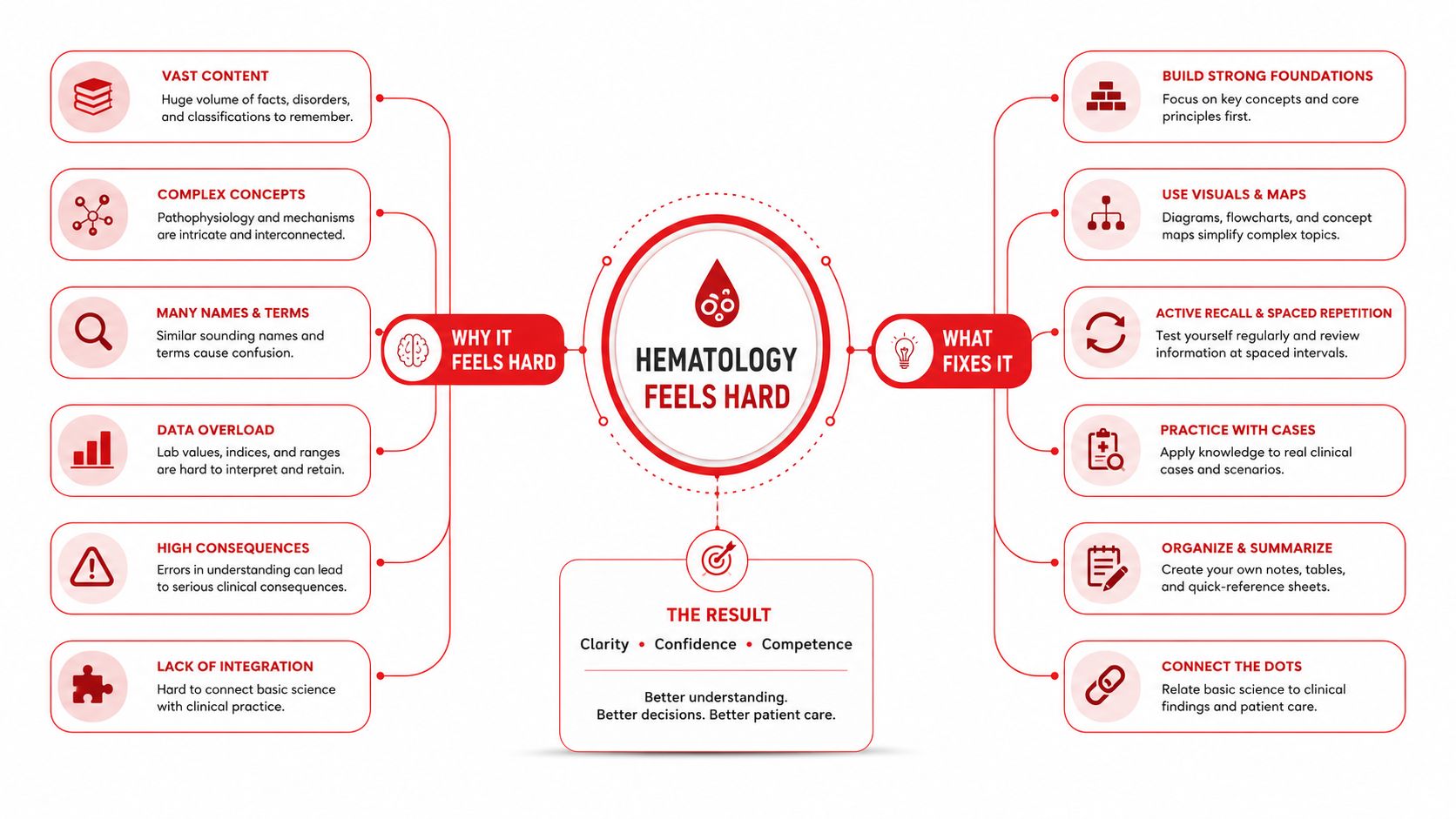
Why Hematology Feels Hard and What Fixes It
A third-year student sees fatigue, bruising, a complete blood count, and a peripheral smear. The instinct is to search memory for a matching disease. That approach breaks down when the stem combines anemia with abnormal platelets, kidney injury, medication exposure, or an unusual smear.
The fix is the Size, Response, Shape, Mechanism framework. Each word answers a different question:
- Size: Is the red blood cell microcytic, normocytic, or macrocytic according to the mean corpuscular volume?
- Response: Is the marrow producing an appropriate number of reticulocytes?
- Shape: What does the peripheral smear reveal, such as schistocytes, spherocytes, hypersegmented neutrophils, or blasts?
- Mechanism: Which test confirms the suspected process, such as iron studies, a direct antiglobulin test, lactate dehydrogenase, haptoglobin, or coagulation testing?
A worked microcytic example
Suppose a hypothetical stem gives fatigue, hemoglobin 8.6 g/dL, mean corpuscular volume 70 fL, a low reticulocyte count, ferritin 5 ng/mL, and increased total iron-binding capacity. Size identifies microcytic anemia. Response shows that the marrow isn't compensating with increased production. Shape may show anisocytosis and hypochromia. Mechanism is iron deficiency, supported by depleted iron stores and increased binding capacity.
The important move isn't recognizing the phrase “microcytic anemia.” It's explaining why thalassemia trait and anemia of inflammation fit less well. Thalassemia generally has a more preserved marrow response and a different iron profile, while inflammation commonly alters iron availability without producing the same depleted-store pattern.
Practical rule: Never stop at the category. Make the next test explain the mechanism.
A structured active recall approach can help you rehearse this sequence until it becomes automatic. Write the four words on a blank page, predict the next finding, and only then check the answer.
How Modern Hematology Was Built
A hematology vignette may begin with a cell count, but the diagnostic task developed through several historical advances: seeing cells, counting them, classifying their appearance, and explaining their mechanism. In 1674, Antonie van Leeuwenhoek described human red blood cells. From the 1770s through the 1820s, William Hewson characterized fibrin, coagulation, and white blood cells. In 1868, Ernst Neumann and Giulio Bizzozero identified bone marrow as the site of hematopoiesis. In 1877, Paul Ehrlich developed blood-cell staining methods that improved leukocyte classification and the diagnosis of blood disease (historical hematology milestones).
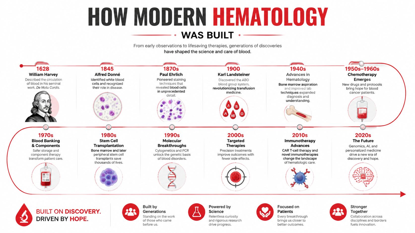
Each advance corresponds to a move in a modern question stem. Cell counters made red-cell indices, including mean corpuscular volume, useful for Size classification. Reticulocyte staining made Response measurable, separating inadequate production from marrow compensation. Romanowsky-type staining made Shape visible through abnormal forms and immature cells. Serologic, immunophenotypic, and molecular tests then supplied increasingly specific evidence for Mechanism.
Treatment developed alongside diagnosis. In 1934, George Whipple, George Minot, and William Murphy demonstrated liver therapy for pernicious anemia, work that became part of a Nobel Prize-winning line of research. In 1949, Linus Pauling characterized sickle hemoglobin, linking a blood disorder to molecular structure, genetics, and protein chemistry.
| Historical advance | Examination skill it supports |
|---|---|
| Cell counting | Classify anemia by red blood cell indices |
| Reticulocyte assessment | Distinguish underproduction from compensation |
| Blood-cell staining | Recognize smear morphology |
| Immunophenotyping | Classify leukemias and lymphomas |
| Molecular and serologic testing | Confirm disease mechanism |
On exam stems, translate each advance into the next question: counting becomes classification, reticulocytes become production assessment, staining becomes morphology recognition, and specialized testing becomes mechanism confirmation.
Normal Blood Cell Physiology You Must Know
Before interpreting disease, know what each blood line is designed to do. Red blood cells transport oxygen, white blood cells coordinate innate and adaptive defense, and platelets provide the first cellular response to vascular injury.
Erythropoietin stimulates erythroid production in the marrow. Mature red cells circulate for about 120 days, while platelets circulate for roughly 7 to 10 days. Platelets arise through fragmentation of megakaryocytes and rapidly adhere, activate, and aggregate after vascular injury, forming an initial platelet plug and helping initiate coagulation (platelet physiology and reference range).
A complete blood count is the foundational screen because it measures red blood cells, hemoglobin, hematocrit, white blood cells, and platelets at the same time. Typical adult reference intervals include 4.6 to 6.2 million red blood cells per microliter in men, 4.2 to 5.4 million in women, and 4,500 to 11,000 white blood cells per microliter (CBC reference intervals). Laboratories vary, so use the provided reference range when one appears in a question.
| Cell Line | Adult Reference Range | Lifespan | Primary Function |
|---|---|---|---|
| Red blood cells | Men, 4.6 to 6.2 million/µL; women, 4.2 to 5.4 million/µL | About 120 days | Oxygen transport |
| White blood cells | 4,500 to 11,000/µL | Varies by lineage | Host defense |
| Platelets | About 150 to 350 ×10^9/L | About 7 to 10 days | Primary hemostasis and inflammation |
Neutrophils are rapid phagocytic responders. Lymphocytes include B cells, T cells, and natural killer cells, so a question about recurrent infections may require you to identify which arm of immunity is affected. Monocytes can become macrophages, while eosinophils and basophils commonly appear in allergy and parasite-related stems.
For studying, a physiology review resource is most useful when you connect normal function to a later decision. Ask what changes first, how the marrow responds, what the smear looks like, and which test confirms the mechanism.
Interpreting the CBC and Peripheral Smear
Hemoglobin 9.1 g/dL, MCV 72 fL, reticulocytes 1.2%, and platelets 245 ×10^9/L. Here is how to move from those values to a mechanism. Begin with Size: the low MCV establishes a microcytic pattern. Then assess Response: the relatively low reticulocyte response suggests that the marrow is not mounting a strong compensatory response. Preserved platelets make a generalized cytopenic process less likely, although the white-cell count and smear still need review.
Use Shape to test the first impression. Anisocytosis and hypochromia support iron-restricted erythropoiesis. Target cells with relatively uniform microcytosis raise thalassemia in the differential. The smear does not replace iron studies or hemoglobin analysis. It identifies the mechanism that deserves confirmation.
| Finding | Pattern | Likely Mechanism |
|---|---|---|
| Schistocytes | Fragmented red cells | Microangiopathic hemolysis |
| Spherocytes | Dense, round red cells without central pallor | Hereditary spherocytosis or warm autoimmune hemolysis |
| Hypersegmented neutrophils | Neutrophils with excessive nuclear segmentation | Megaloblastic hematopoiesis |
| Blasts | Immature precursor cells | Acute leukemia or another marrow process |
| Rouleaux | Stacked red cells | Increased plasma proteins, including in plasma-cell disorders |
The final step is Mechanism. Ask what process could produce the size, response, and shape together, then choose the test that separates the leading possibilities. Add Timing and Trend: an abrupt change suggests a different process from a longstanding stable pattern, and serial CBCs can reveal progression before a single result does.
A reusable CBC vignette sequence
- Syndrome: Is the dominant problem anemia, infection, bleeding, thrombosis, pancytopenia, or a mass-related process?
- Numbers: Which cell line is abnormal, and is the change isolated or multilineage?
- Size: What does the MCV suggest?
- Response: Is reticulocyte production appropriate?
- Shape: Which smear finding changes the differential?
- Mechanism: What confirmatory test should come next?
- Timing and Trend: Did the abnormality appear suddenly, persist, or evolve across serial results?
A lab-value review sheet can help with reference-range recall, but question review should connect each result to the next diagnostic decision. Recognizing schistocytes is only the beginning. Connect them to hemolysis, possible thrombocytopenia, kidney injury, and the need for urgent evaluation. That connection turns a memorized shape into clinical reasoning.
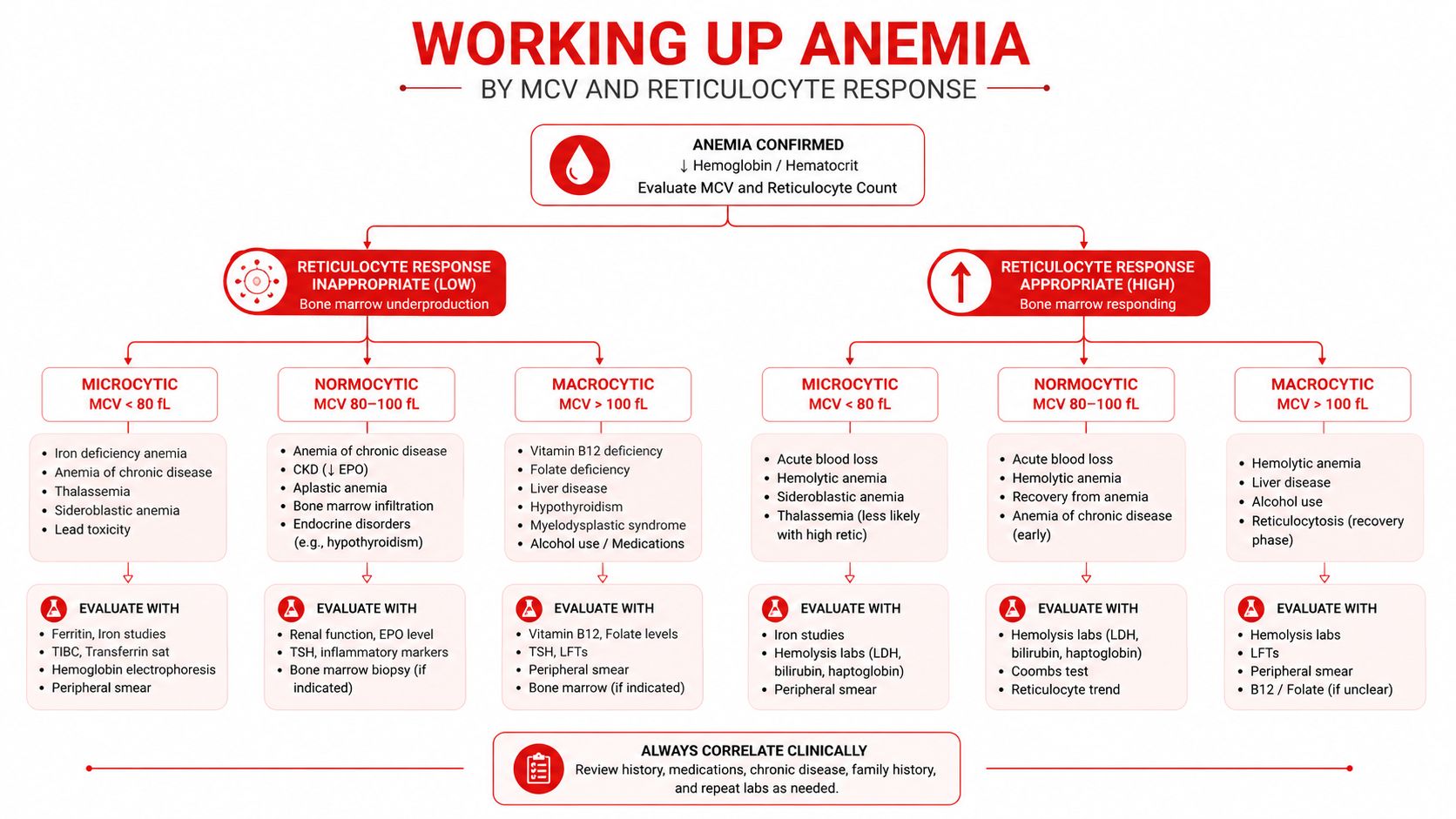
Working Up Anemia by MCV and Reticulocyte Response
Anemia is defined in adults by hemoglobin below 13.6 g/dL in adult males and below 12 g/dL in adult females. One clinical reference also identifies hematocrit below 40% in males and below 37% in females, with red blood cell counts below 4.5 million/mcL in males and below 4 million/mcL in females (adult anemia thresholds).
Start with mean corpuscular volume:
- Microcytic: below 80 fL
- Normocytic: 80 to 100 fL
- Macrocytic: above 100 fL
Then ask whether the marrow response is appropriate. A reticulocyte production index corrects the reticulocyte percentage for the degree of anemia and the shortened survival of circulating reticulocytes. A low corrected response suggests underproduction. A high response supports blood loss or hemolysis, provided the marrow has the capacity to respond.
Use iron studies and hemolysis markers deliberately
Microcytic anemia with low reticulocytes calls for iron studies. Ferritin below 30 ng/mL is highly sensitive for iron deficiency in the general population, while the American Gastroenterological Association recommends a cutoff below 45 ng/mL when evaluating gastrointestinal causes (ferritin thresholds). Low ferritin and increased total iron-binding capacity support iron deficiency. Anemia of inflammation more often has reduced iron availability with a normal or increased ferritin, while thalassemia trait suggests hemoglobin analysis, including hemoglobin A2 assessment. Sideroblastic processes require a different mechanism-focused evaluation.
Macrocytosis requires separation of megaloblastic from nonmegaloblastic causes. Vitamin B12 and folate testing, methylmalonic acid, homocysteine, medication history, alcohol exposure, liver disease, and thyroid disease help organize the differential. Hypersegmented neutrophils make a megaloblastic process more likely, but they don't identify the specific vitamin deficiency by themselves.
Normocytic anemia with reticulocytosis, increased lactate dehydrogenase, increased indirect bilirubin, and low haptoglobin points toward hemolysis. A direct antiglobulin test can then distinguish immune from nonimmune hemolysis. Normocytic anemia with low reticulocytes instead favors impaired production, inflammation, kidney-related erythropoietin deficiency, or aplastic marrow failure.
Reasoning sequence: Size first, response second, shape third, mechanism last.
For a rapid workup order under exam pressure, use this sequence:
- Confirm anemia and classify the mean corpuscular volume.
- Check the reticulocyte response.
- Review the smear.
- Order iron studies for microcytosis, vitamin studies for macrocytosis, or hemolysis markers for reticulocytosis.
- Choose targeted testing based on the mechanism rather than ordering every test at once.
Platelet Versus Coagulation Disorders
The bleeding pattern often tells you which hemostatic system failed before you interpret the laboratory values. Primary hemostasis, involving vessels and platelets, produces mucocutaneous bleeding. Secondary hemostasis, involving coagulation factors, produces bleeding into deeper tissues.
Petechiae, epistaxis, gingival bleeding, and heavy menstrual bleeding suggest thrombocytopenia or platelet dysfunction. Hemarthroses, deep muscle hematomas, and delayed postoperative bleeding suggest factor deficiency. Platelets normally circulate at about 150 to 350 ×10^9/L and form the initial plug after vascular injury (platelet function and counts).
| Disorder | Bleeding Pattern | PT | PTT | Platelets | Confirmatory Test |
|---|---|---|---|---|---|
| Immune thrombocytopenia | Petechiae and mucosal bleeding | Normal | Normal | Low | Diagnosis of exclusion |
| Hemophilia A or B | Deep bleeding and hemarthroses | Normal | Prolonged | Normal | Factor assay |
| von Willebrand disease | Mucocutaneous bleeding | Usually normal | Normal or prolonged | Usually normal | vWF antigen and activity testing |
| Disseminated intravascular coagulation | Mucosal and deep bleeding | Prolonged | Prolonged | Low | Fibrinogen, D-dimer, smear, clinical context |
| Warfarin effect | Variable bleeding | Prolonged | May be normal or prolonged | Usually normal | Medication review and coagulation testing |
Let the laboratory pattern refine the differential
An isolated low platelet count with normal prothrombin time and partial thromboplastin time suggests a platelet-focused process such as immune thrombocytopenia, but thrombotic thrombocytopenic purpura and other emergencies require attention to hemolysis, neurologic findings, renal injury, fever, and the smear. Schistocytes change the urgency because they indicate red-cell fragmentation rather than isolated immune platelet destruction.
An isolated prolonged partial thromboplastin time can reflect hemophilia, heparin exposure, or an inhibitor. A mixing study asks whether normal plasma corrects the abnormality. Correction supports a factor deficiency, while failure to correct suggests an inhibitor. Factor assays and von Willebrand testing then define the mechanism.
Combined prothrombin-time and partial-thromboplastin-time prolongation raises warfarin effect, vitamin K deficiency, liver disease, or disseminated intravascular coagulation. Heparin-induced thrombocytopenia requires a medication timeline, platelet trend, thrombosis assessment, and appropriate antibody testing rather than reflexively labeling every postoperative thrombocytopenia as immune thrombocytopenia.
A pharmacology review resource can help connect anticoagulant mechanisms with coagulation patterns. On questions, first identify the bleeding compartment, then the abnormal laboratory pathway, then the confirmatory test.
Transfusion Medicine Anticoagulation and Malignancies
Transfusion questions reward prioritization. First decide whether the patient needs immediate stabilization. Then identify the blood product, compatibility issue, or reversal agent that addresses the active danger.
Packed red blood cell decisions depend on clinical context, symptoms, ongoing loss, and institutional guidance. Platelet transfusion decisions also depend on bleeding, procedures, and the cause of thrombocytopenia. Don't treat a laboratory value as a substitute for the patient's hemodynamic status.
For product selection, remember the underlying purpose:
- Red cells: Improve oxygen-carrying capacity.
- Platelets: Support primary hemostasis.
- Plasma: Replace multiple coagulation factors.
- Cryoprecipitate: Replace fibrinogen and selected concentrated proteins.
Reactions and anticoagulant reversal
An acute hemolytic transfusion reaction suggests incompatible red-cell exposure. Stop the transfusion, maintain appropriate access, notify the blood bank, and evaluate the patient according to institutional emergency protocols. Fever, hypoxemia, pulmonary edema, and hypotension require differentiation among acute hemolysis, transfusion-related acute lung injury, transfusion-associated circulatory overload, and other causes.
| Agent | Reversal | Notes |
|---|---|---|
| Warfarin | Vitamin K plus prothrombin complex concentrate for serious bleeding | Corrects vitamin K-dependent factor deficiency |
| Dabigatran | Idarucizumab | Direct thrombin inhibitor reversal |
| Factor Xa inhibitors | Andexanet alfa when indicated by current guidance and availability | Direct factor Xa inhibitor reversal |
| Unfractionated heparin | Protamine | Neutralizes heparin activity |
Malignancy patterns and emergencies
Acute leukemia questions often combine constitutional symptoms, cytopenias, blasts, and infection or bleeding. For acute myeloid leukemia, the traditional World Health Organization morphologic threshold is at least 20% blasts in bone marrow or peripheral blood, although WHO 2022 rules remove that requirement for some entities with defining genetic abnormalities (acute myeloid leukemia diagnostic criteria).
Lymphoma questions require attention to distribution, constitutional symptoms, immunophenotype, and staging logic. Myeloma questions use the CRAB pattern, hypercalcemia, renal dysfunction, anemia, and bone disease, along with monoclonal protein findings. Hyperleukocytosis, tumor lysis syndrome, and Richter transformation are time-sensitive red flags. In each case, identify the immediate threat before naming the definitive therapy.
Study Resources and Next Steps for Hematology Prep
Use resources for different jobs rather than asking one source to teach everything. A foundational text such as Hoffman or Wintrobe can deepen physiology and mechanisms. Pathoma chapter 5 and the hematology section of First Aid can support rapid pattern recognition. A question bank such as UWorld, AMBOSS, or TrueLearn can provide original practice vignettes, but none should replace careful review of official exam information or your school's curriculum.
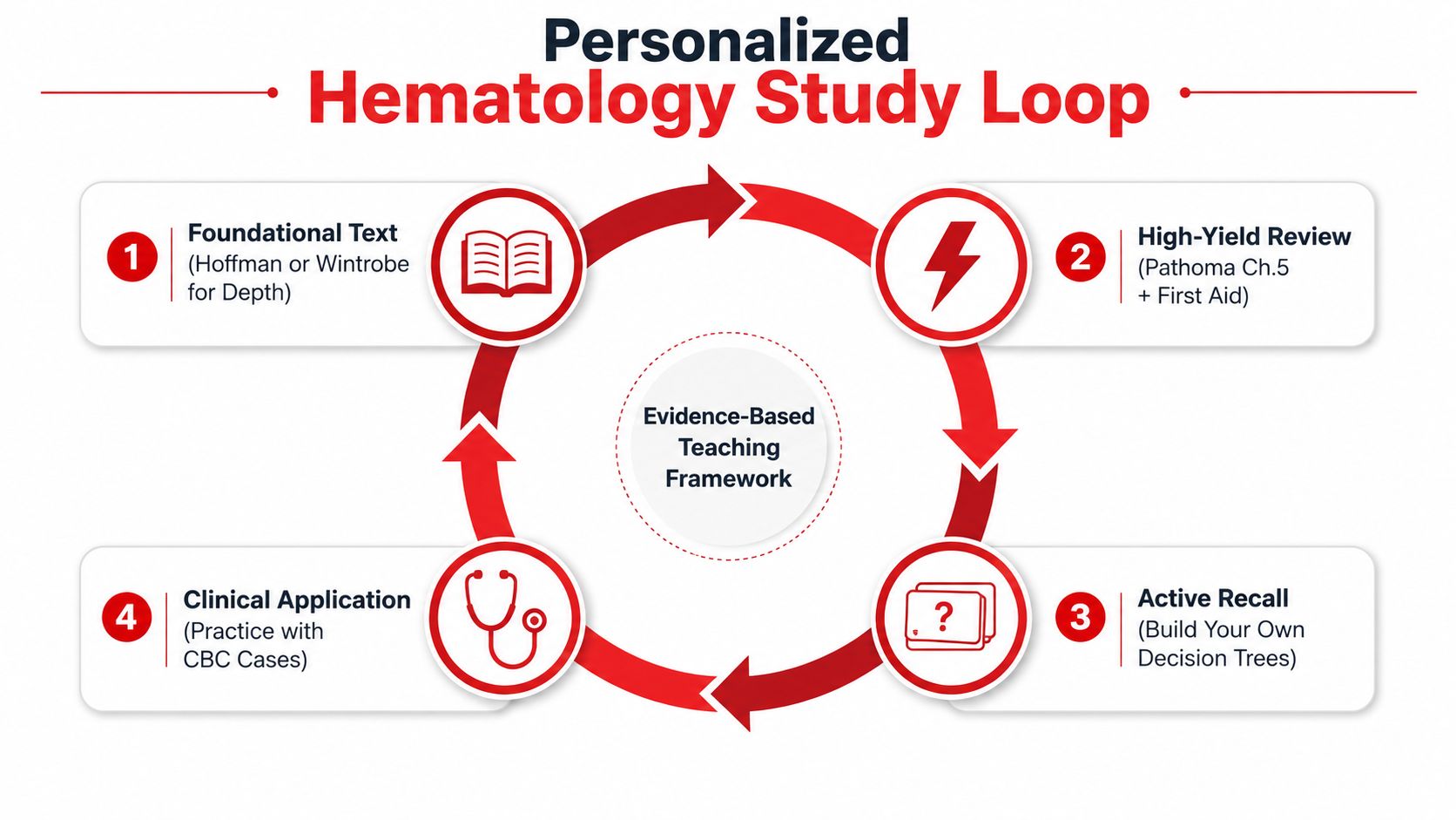
Map each resource to the four-part framework:
- Foundational reading: Repair physiology and mechanisms.
- High-yield review: Organize Size and Shape patterns.
- Question practice: Test Response and next-best-test decisions.
- Smear review: Link morphology to Mechanism.
A practical 14-day plan
| Days | Main task | Output |
|---|---|---|
| 1 to 3 | Review red-cell physiology and anemia by mean corpuscular volume | One anemia decision tree |
| 4 to 6 | Complete mixed anemia questions and classify every miss | Error log by Size, Response, Shape, or Mechanism |
| 7 to 8 | Review platelets, coagulation, and anticoagulants | Bleeding-pattern comparison |
| 9 to 10 | Study transfusion reactions and blood products | Immediate-stabilization checklist |
| 11 to 12 | Review leukemia, lymphoma, and myeloma | Malignancy pattern table |
| 13 | Complete an unseen mixed hematology block under timed conditions | Accuracy and pacing audit |
| 14 | Revisit errors and teach the framework aloud | Final one-page summary |
Use spaced repetition for medical learning to revisit missed mechanisms rather than rereading entire chapters. Common traps include relying on mean corpuscular volume alone, ignoring the smear, calling every thrombocytopenia immune thrombocytopenia, and confusing thrombotic thrombocytopenic purpura with a simple platelet disorder.
Your checkpoint should answer four questions:
- Syndrome: What clinical problem is present?
- Sequence: Did I use Size, Response, Shape, and Mechanism in order?
- Distractors: Why are the two closest alternatives less likely?
- Action: What requires immediate stabilization, and what confirms the diagnosis?
For transparent progress measurement, record timed accuracy on an unseen mixed hematology block, tag each error as knowledge, reasoning, misread, or pacing, then reassess with a different unseen block after spaced review. If real internal data are ever reported, include the sample size, median change, spread, time interval, and question source rather than presenting an unsupported average.
Clinical information in this guide is for examination education, not diagnosis or medical treatment. Laboratory reference ranges and clinical protocols vary, so real patient care requires current institutional guidance and supervision.
If you want individualized help applying this framework to USMLE or COMLEX hematology questions, Ace Med Boards offers one-on-one tutoring, question analysis, and study planning based on the learner's documented needs. You can schedule a free consultation to review your current approach and decide whether targeted support fits your preparation.