
You're staring at a chest X-ray during a Shelf exam, recognizing that something looks abnormal but not knowing where to begin. Radiology becomes much more manageable when you stop treating images as isolated pictures and start reading them as structured clinical evidence. For USMLE, COMLEX-USA, and Shelf preparation, the goal is to identify the modality, confirm the anatomy, apply a consistent search pattern, and connect the finding to the patient's presentation.
Radiology Basics for Medical Exams
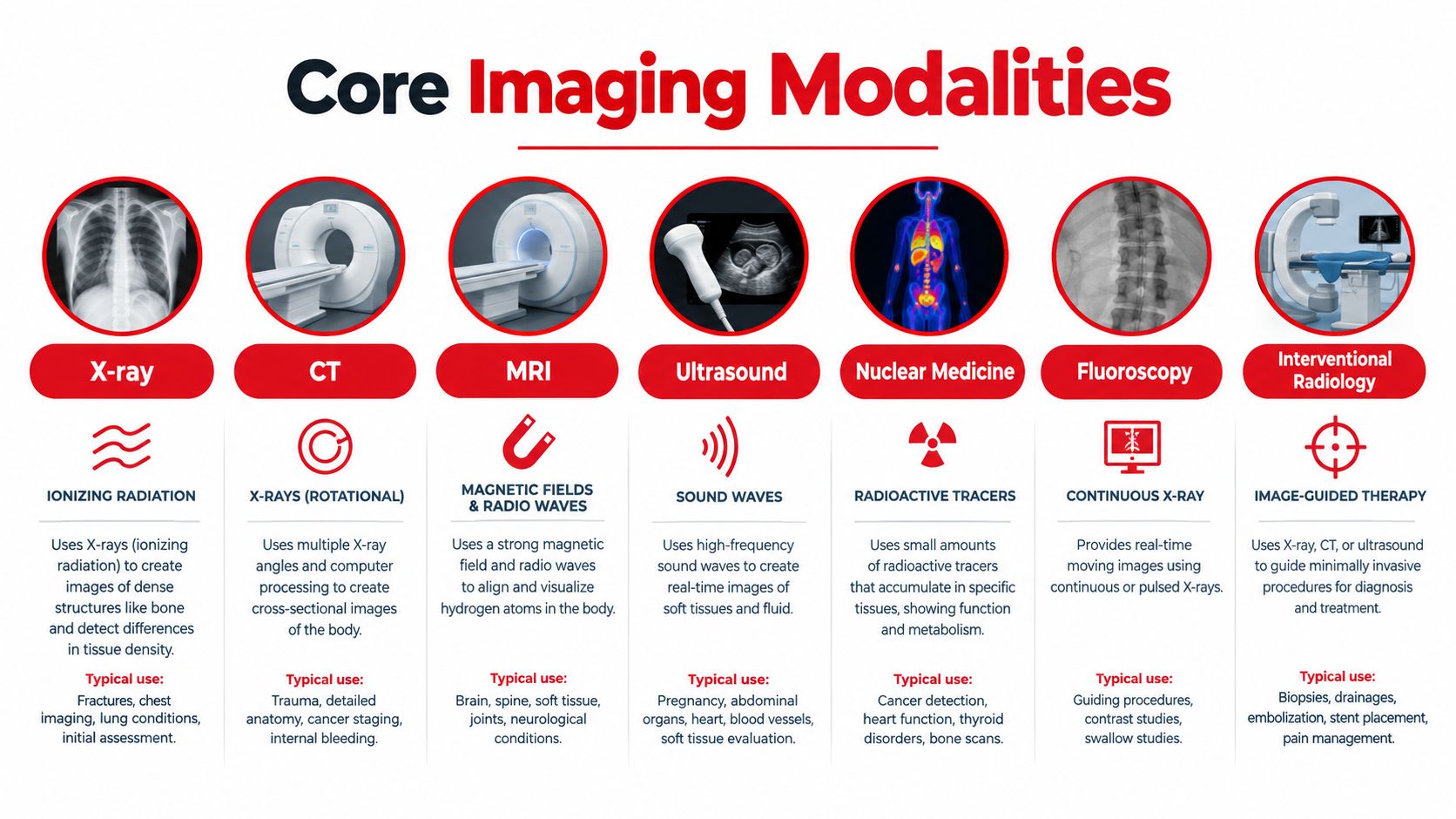
Radiology is the medical specialty that uses imaging to diagnose disease and, in some settings, guide treatment. Its scope includes plain X-rays, computed tomography (CT), magnetic resonance imaging (MRI), ultrasound, nuclear medicine, fluoroscopy, and image-guided procedures performed through interventional radiology.

Radiology matters on medical exams because imaging findings appear inside questions about many organ systems, not only in questions labeled “radiology.” A patient with sudden dyspnea may have a pneumothorax on a chest radiograph. A patient with right lower-quadrant pain may have appendicitis on CT. A patient with focal neurologic deficits may need an imaging-based distinction between hemorrhage and ischemia. The image is often the decisive piece of evidence after the stem has already established the clinical context.
A short history with a lasting exam lesson
Radiology began with Wilhelm Conrad Röntgen's discovery of X-rays on November 8, 1895. Within weeks, practical medical applications were underway, and on December 22, 1895, Röntgen produced the first radiograph, an image of his wife's hand. That milestone changed medicine by allowing physicians to see inside the body without surgery. Historical development continued rapidly, with early contrast studies in the late 1890s, cerebral angiography in 1927, and cardiac angiography in 1929. A historical review of radiology's development places these advances in context.
For exam purposes, think in a sequence:
- What modality am I viewing?
- Which structures should normally be visible?
- What is the dominant abnormal pattern?
- Which diagnosis best fits both the image and the clinical stem?
Practical rule: Don't name the disease before you've described the finding. “A unilateral pleural line with absent peripheral lung markings” is stronger reasoning than “probably pneumothorax.”
Core Imaging Modalities Explained
Each modality answers a different question. Instead of memorizing long lists, match the technology to the tissue, urgency, and clinical problem.
| Modality | How it works in simple terms | Strong exam applications | Radiation |
|---|---|---|---|
| X-ray | Uses ionizing radiation to create a two-dimensional projection | Fractures, chest pathology, bowel gas patterns | Yes |
| CT | Combines multiple X-ray projections into cross-sectional images | Trauma, acute bleeding, stones, abdominal emergencies | Yes |
| MRI | Uses magnetic fields and radiofrequency energy to characterize tissues | Brain, spinal cord, joints, soft tissue | No ionizing radiation |
| Ultrasound | Sends sound waves into the body and analyzes returning echoes | Gallbladder, pregnancy, vascular flow, bedside assessment | No ionizing radiation |
| Nuclear medicine | Tracks a radiolabeled substance according to physiologic activity | Bone activity, thyroid function, perfusion and metabolism | Uses radioactive material |
| Fluoroscopy | Produces moving X-ray images in real time | Swallow studies, contrast examinations, procedural guidance | Yes |
| Interventional radiology | Uses imaging to guide minimally invasive diagnosis or treatment | Biopsy, drainage, vascular procedures, embolization | Often uses imaging, commonly including fluoroscopy or CT |
X-ray and CT
An X-ray is like looking at a shadow map. Dense materials such as bone appear bright, while air appears dark. The limitation is overlap. A frontal chest radiograph compresses three-dimensional anatomy into one projection, so position, rotation, and additional views matter.
CT is closer to slicing a loaf of bread and examining each slice separately. It reduces anatomic overlap and provides cross-sectional detail, which makes it valuable in trauma and acute abdominal disease. Effective dose varies by protocol. One comparative summary lists a chest CT at about 7.0 mSv, an abdomen CT at about 6.8 mSv, and an abdomen and pelvis CT at about 14.0 mSv, compared with about 0.1 mSv for a chest radiograph and about 0.001 mSv for an extremity X-ray. A clinical radiation-safety summary explains why justification and protocol selection matter.
For a visual overview of how these tools differ, review these postmortem CT scan examples, then practice applying modality selection with this chest X-ray interpretation guide.
MRI, ultrasound, and functional imaging
MRI excels at soft-tissue contrast. On exams, associate it with the brain, spinal cord, ligaments, cartilage, and marrow. T1-weighted and T2-weighted sequences are common testing targets, but the clinical stem usually tells you what to prioritize. MRI is powerful but may be less suitable when the patient is unstable or cannot tolerate a lengthy examination.
Ultrasound depends on sound-wave reflection. It's portable, avoids ionizing radiation, and can show motion or blood flow with Doppler. High-yield associations include gallstones, hydronephrosis, deep venous thrombosis evaluation, and pregnancy-related imaging.
Nuclear medicine emphasizes function rather than anatomy alone. Fluoroscopy shows motion in real time, while interventional radiology turns imaging into procedural guidance. The exam question may test not only recognition, but also why one modality is preferable for the clinical situation.
Normal Anatomy and Common Pathologies by System
A reliable image reader compares expected anatomy with the pattern that is present. Start with the system named in the stem, but keep adjacent structures in view because many abnormalities cross anatomic boundaries.
Chest
| Normal expectation | Common abnormal pattern |
|---|---|
| Lung markings extend toward the periphery | A visible pleural line with absent peripheral markings suggests pneumothorax |
| Cardiac borders are distinguishable when not obscured | Loss of a border can indicate adjacent consolidation, the silhouette sign |
| Diaphragms are smooth, with the right often slightly higher | Free subdiaphragmatic air raises concern for perforation |
| Mediastinum is assessed for width and contour | Widening may suggest a vascular or mediastinal process, interpreted with the clinical stem |
Pneumonia often appears as focal or lobar air-space opacity, but the distribution and silhouette sign help localize it. Don't call every white region pneumonia. Ask whether the opacity is truly within the lung, whether there's volume loss, and whether the patient's symptoms support infection.
Abdomen
- Normal bowel: Gas may appear in the stomach and bowel, but the pattern should be assessed for distribution and dilation.
- Obstruction: Dilated loops and air-fluid levels support a mechanical or functional obstruction, with the transition point and clinical context guiding interpretation.
- Appendicitis: CT may show an enlarged, inflamed appendix with surrounding inflammatory change. Read the entire right lower quadrant rather than stopping after finding nonspecific pain-related changes.
- Free air: Gas outside the bowel lumen can indicate perforation and requires urgent clinical correlation.
For a focused review of phases, anatomy, and common findings, use this abdominal CT reading guide.
Musculoskeletal and neurologic imaging
In musculoskeletal questions, trace the cortex continuously. A subtle cortical break, abnormal alignment, or joint-space change may matter more than dramatic soft-tissue swelling. On MRI, identify the structure named in the stem first, then evaluate signal and continuity.
Neurologic imaging rewards localization. On head CT, acute blood is typically hyperdense, while ischemic changes may be subtle early. MRI is often emphasized for soft-tissue and neurologic detail. Always match the imaging pattern to onset, focal deficits, trauma, infection risk, or malignancy clues.
Read the image in the same order every time. A consistent search pattern protects you from anchoring on the first abnormality you notice.
Image Interpretation Frameworks and Case Walkthroughs
A framework helps you find abnormalities before the answer choices start pulling your attention in different directions. The framework isn't a substitute for anatomy. It's a guardrail against omission.
ABCDE for chest X-rays
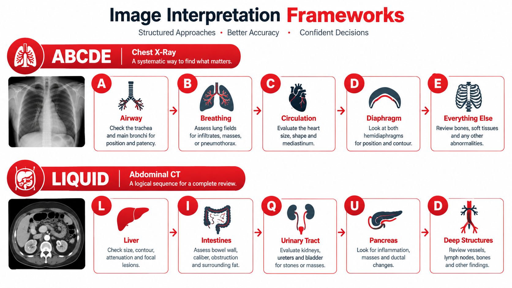
Use ABCDE as a practical sequence:
- A, Airway: Check tracheal position and major airway alignment.
- B, Breathing: Compare lung fields, pleural spaces, and vascular markings.
- C, Circulation: Review heart size, mediastinal contours, and pulmonary vessels.
- D, Diaphragm: Look for free air, pleural fluid, and costophrenic-angle changes.
- E, Everything else: Inspect bones, soft tissues, lines, tubes, and external artifacts.
Suppose an original practice vignette describes fever, productive cough, and focal crackles. You see a focal air-space opacity near a recognizable cardiac border. Under ABCDE, the airway is midline, the opposite lung is clear, the heart isn't globally enlarged, and there's no pleural line. The local opacity plus the clinical presentation supports pneumonia, while the silhouette sign helps with localization.
The common error is premature closure. A student sees opacity and stops. A stronger reader checks for pleural effusion, pneumothorax, collapse, and device-related complications before selecting an answer.
LIQUID for abdominal CT
For abdominal CT, LIQUID can organize a region-by-region search:
- L, Liver: Review parenchyma, vessels, and biliary structures.
- I, Intestines: Follow bowel caliber, wall, enhancement, and surrounding fat.
- Q, Quadrants: Compare the upper and lower quadrants systematically.
- U, Urinary tract: Inspect kidneys, ureters, and bladder.
- I, Inflammation and fluid: Look for fat stranding, collections, and free fluid.
- D, Devices and bones: Check lines, drains, hardware, and visible osseous structures.
In an original obstruction-style case, a patient has vomiting, abdominal distension, and cramping. CT shows dilated proximal bowel with a transition to decompressed distal bowel. LIQUID directs you to confirm the bowel pattern, search for an obstructing lesion or twisting, and assess for ischemic clues such as abnormal wall enhancement or concerning free fluid.
Avoid the opposite mistake, overcalling normal variants. Mild dependent fluid, anatomic asymmetry, or postoperative changes may be incidental. Clinical reasoning requires you to ask whether the imaging finding explains the patient's symptoms and whether the stem supplies evidence of complications. This clinical reasoning resource can help connect imaging observations to diagnosis and next steps.
Study Strategies and Resources for Exam Success
Radiology review works best when you practice recognition and reasoning together. Looking at dozens of images without explaining your interpretation creates familiarity, but not necessarily retrieval under pressure. A question bank such as UWorld, official National Board of Medical Examiners (NBME) materials for relevant examinations, and a focused radiology review book can serve different purposes. Use each according to its role, and remember that third-party resources aren't affiliated with the examination organizations unless explicitly stated.
A practical weekly rhythm
A sustainable schedule might look like this:
- Early week: Review one modality or organ system, then annotate a small set of representative images.
- Middle of week: Complete timed questions and record the exact visual clue that determined each answer.
- Later week: Revisit missed images without looking at the explanation, then explain the diagnosis aloud.
- End of week: Mix chest, abdominal, neurologic, and musculoskeletal cases to test transfer rather than memorization.
Use spaced repetition for image features, not just disease names. A flashcard should ask, “What finding distinguishes this from its closest mimic?” rather than only, “What is the diagnosis?” For additional techniques, this guide on how to improve retention with active recall offers a useful study-method comparison.
Readiness checklist
Before an exam, confirm that you can:
- Name the modality: Explain what the modality shows well and what it misses.
- Use a sequence: Apply ABCDE to chest radiographs and LIQUID to abdominal CT practice.
- Describe before diagnosing: State location, density or signal, distribution, and associated findings.
- Separate pattern from context: Use the stem to prioritize, not to force an image to fit.
- Review errors actively: Write why your chosen answer was wrong and what visual clue you overlooked.
Keep a short “image error log.” Divide mistakes into recognition errors, anatomy errors, framework omissions, and clinical reasoning errors. Ace Med Boards offers individualized tutoring for USMLE, COMLEX-USA, and selected Shelf preparation, so learners who need external structure can compare that option with independent study and official practice materials through the medical student study tips resource.
Radiology Workforce and Access Considerations
Radiology questions increasingly sit within a healthcare system that must balance growing imaging demand, limited expertise, appropriate utilization, and unequal access. The American College of Radiology (ACR) reports that the United States has about 1,500 fewer radiologists than needed and could face a shortage of roughly 3,100 soon. The same update reports that only around 30% of radiologists use artificial intelligence in routine clinical workflows, so AI adoption hasn't resolved the staffing problem. The ACR workforce update provides the cited figures and context.
Access also varies by geography and resources. Rural communities may have fewer imaging options and longer waits, while Asia-Oceania radiology leaders describe substantial urban-rural disparities. Staffing data from 2025 reported vacancy rates near record highs in several modalities, including 19.4% for CT and 17.4% for MRI. The ASRT staffing and workplace survey helps explain why new machines alone don't guarantee timely imaging.
Why appropriateness appears on exams
Exams may test whether you choose an imaging study that answers the clinical question without unnecessary exposure or delay. The ACR Appropriateness Criteria covered 233 diagnostic imaging and interventional radiology topics by 2023, with more than 1,100 variants and about 3,000 clinical scenarios for diagnostic imaging topics. ACR's clinical decision-support overview describes the breadth of the tool.
The rating scale runs from 1 to 9. Ratings of 1 to 3 mean a study is usually not appropriate, 4 to 6 mean it may be appropriate, and 7 to 9 mean it is usually appropriate. This AAFP explanation of the criteria provides the scale.
Training capacity is also competitive. The 2026 NRMP Main Residency Match listed 877 diagnostic radiology positions, including 354 U.S. MD seniors and 299 non-U.S. citizen international medical graduates in the specialty match pool. The NRMP 2026 results report contains those figures. A Radiology journal analysis reported 1,356 available radiology positions in 2023 across the specified diagnostic and integrated programs, with a 67.2% crude match rate for all applicant types.
For applicants considering the specialty, this radiology residency application guide addresses application planning without treating one score or one setback as a final verdict. For referral workflow context, readers can also review this guide to radiology referrals for GPs.
Ace Med Boards offers personalized online tutoring for USMLE, COMLEX-USA, and selected Shelf exams, including structured image interpretation, question-analysis practice, and targeted study planning. If radiology questions are a recurring weakness, visit Ace Med Boards to learn about available support and decide whether a free consultation fits your preparation needs.