Chapman points are small, firm, tender fascial nodules taught as organ-associated reflex points. For the Osteopathic Medical Licensing Examination of the United States (COMLEX-USA), the learning task is to recognize the location-to-organ pairing, not treat a point as a standalone diagnosis.
What happens when a student recognizes the organ but confuses the side, rib, or posterior landmark? The answer can still be wrong. Effective Chapman points COMLEX preparation combines an anatomical lookup with retrieval practice and careful reading of what the vignette asks.
Ace Med Boards Editorial Team
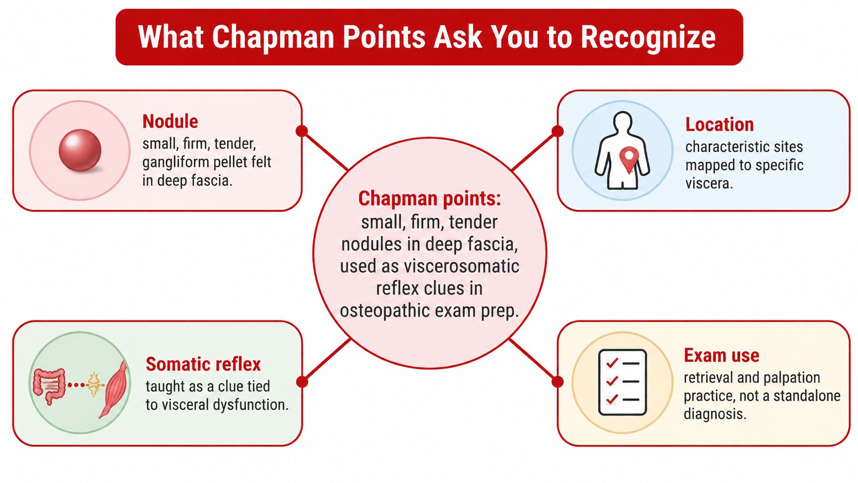
What Chapman Points Ask You to Recognize
What is a COMLEX vignette asking when it describes a tender nodule at a named landmark? Usually, the task is to identify the site, recall its traditional organ pairing, and judge whether the clinical presentation fits. The finding is a clue to interpret, not a diagnosis by itself.
Frank Chapman, DO, described these points as neuro-lymphatic reflex points, according to the NCBI Bookshelf clinical overview of Chapman points. Later osteopathic literature developed the concept of palpable nodules associated with visceral dysfunction through segmental relationships. That history explains the terminology, while the proposed reflex mechanism should not be treated as established diagnostic accuracy.
The language behind the finding
In osteopathic manipulative medicine, students also learn tissue texture change, asymmetry, restriction of motion, and tenderness, often abbreviated TART. These terms describe somatic findings, but they do not all identify a specific Chapman point.
Tissue texture change refers to altered consistency, such as increased firmness. Asymmetry is a difference between corresponding structures or sides; restriction is reduced motion; tenderness is discomfort elicited during examination. Keep the terms distinct when a question gives several findings.
Key Takeaways
- Pairings support recognition. Use the named landmark and side to distinguish closely related answer choices.
- The mechanism remains unsettled. A traditional reflex explanation does not establish a confirmed physiological mechanism.
- A point does not diagnose disease. Interpret the finding alongside the broader clinical picture.
- Red flags take priority. Objective evidence and concerning symptoms outweigh an isolated reflex association.
Treat these associations as examination aids, not medical advice or a treatment guide. The charts that follow are teaching references, not independently clinician-reviewed diagnostic maps. Match the locations to the osteopathic atlas and syllabus used in your course.
Anterior Chapman Points and Organ Pairings
Anterior Chapman points commonly taught for COMLEX
Every row represents a traditional examination association, not a measured diagnostic coordinate. The common-swap column identifies a recall trap, not an alternative diagnosis.
| Organ or region | Side | Anterior teaching location | Common swap |
|---|---|---|---|
| Middle ear | Bilateral | Superior to the medial clavicle | Sinus point just below the clavicle |
| Sinuses | Bilateral | Inferior to the medial clavicle | Middle-ear point above the clavicle |
| Pharynx | Midline | Anterior first rib near the sternum | Tonsil in the first intercostal space |
| Tonsils | Bilateral | First intercostal space at the sternal border | Pharynx on the first rib |
| Tongue | Bilateral | Second rib costochondral junction | Esophagus in the second interspace |
| Esophagus, thyroid, myocardium, bronchi | Bilateral or midline teaching grouping | Second intercostal space near the sternum | Upper lung in the third interspace |
| Upper lung | Bilateral | Third intercostal space near the sternum | Lower lung in the fourth interspace |
| Lower lung | Bilateral | Fourth intercostal space near the sternum | Upper lung in the third interspace |
| Stomach acidity | Left | Left fifth intercostal space | Liver on the right fifth to sixth |
| Stomach peristalsis | Left | Left sixth intercostal space | Spleen on the left seventh |
| Liver | Right | Right fifth to sixth intercostal spaces | Gallbladder more lateral at the sixth |
| Gallbladder | Right | Right sixth intercostal space, midclavicular line | Liver closer to the sternum |
| Pancreas | Right | Right seventh intercostal space | Spleen on the left seventh |
| Spleen | Left | Left seventh intercostal space | Stomach on the left fifth or sixth |
| Small intestine | Bilateral | Eighth to tenth intercostal spaces near the costochondral junctions | Colon on the thigh |
| Appendix | Right | Tip of the right twelfth rib | Cecum on the proximal right thigh |
| Adrenal | Bilateral | About two inches superior and one inch lateral to the umbilicus | Kidney, which sits closer to the umbilicus |
| Kidney | Bilateral | About one inch superior and one inch lateral to the umbilicus | Adrenal, which sits higher |
| Bladder | Midline | Periumbilical fascia | Urethra at the pubic symphysis |
| Urethra | Midline | Superior pubic symphysis | Bladder at the umbilicus |
| Cecum | Right | Proximal right anterior iliotibial band | Appendix at the twelfth-rib tip |
| Ascending colon and hepatic flexure | Right | Right anterior iliotibial band, progressing distally | Cecum at the proximal end |
| Sigmoid colon | Left | Proximal left anterior iliotibial band | Descending colon farther down the left band |
| Descending colon and splenic flexure | Left | Left anterior iliotibial band, mid to distal | Sigmoid at the proximal left band |
Read landmarks before organ names
The anterior map becomes easier to organize when students separate ribs from intercostal spaces. A point on a rib isn't the same landmark as a point in the space below it. That distinction matters particularly in the upper chest, where several teaching associations occupy a compact area.
The grouped esophagus, thyroid, myocardium, and bronchi row is a regional memory aid. It shouldn't suggest that every structure has an identical, universally agreed coordinate. A stem that supplies a more exact location should be interpreted using the convention taught in the assigned course resource.
Landmark rule: Read the structure first, then the side, then the organ choices. A familiar organ name shouldn't pull attention away from the actual location.
Separate side traps from neighbor traps
The upper abdominal associations are especially vulnerable to right-left reversals. Stomach pairings belong on the left; liver and gallbladder pairings belong on the right. Pancreas and spleen form another useful contrast because the teaching level is similar while the side changes.
The peri-umbilical cluster requires a different strategy. Students should sketch the umbilicus and place the adrenal site above the kidney site, rather than memorizing disconnected measurements. The bladder and urethra then remain separate by reference landmark: umbilical versus pubic.
For colon associations, the anterior iliotibial band is the organizing structure. Cecum and sigmoid occupy opposite proximal thigh associations and are easy to swap when a student remembers only “colon on the thigh.” Ovaries and testes are traditionally placed anteriorly near the pubic tubercle, while the uterus is associated with the inferior pubic ramus region.
Exact point conventions differ across teaching resources. Before incorporating these locations into a permanent flashcard deck, students should compare them with the school syllabus rather than assume that a broad chart resolves every anatomical detail.
Posterior Chapman Points by Spinal Level
The posterior map works best as a top-to-bottom anatomical scan. Cervical articular pillars organize head-and-neck associations; thoracic landmarks organize chest and abdominal associations; lumbar and sacral regions organize lower abdominal and pelvic associations.
Posterior Chapman points commonly taught for COMLEX
Each row remains a traditional organ association with limited diagnostic evidence. Broad level labels are recall shorthand, not instructions to palpate a spinous process or diagnose an organ disorder.
| Organ or region | Side | Posterior teaching location | Anterior partner to recall |
|---|---|---|---|
| Middle ear | Bilateral | C1 articular pillar | Above the medial clavicle |
| Sinuses, pharynx, larynx, tongue | Bilateral | C2 articular pillar region | Clavicle, first rib, or second rib |
| Tonsils | Bilateral | C1 region, often grouped with atlas | First intercostal space |
| Esophagus, thyroid, bronchi, myocardium | Bilateral | T2 to T3 intertransverse space | Second intercostal space |
| Upper lung | Bilateral | T3 intertransverse space | Third intercostal space |
| Lower lung | Bilateral | T4 intertransverse space | Fourth intercostal space |
| Stomach | Left | Left T5 to T6 | Left fifth and sixth intercostal spaces |
| Liver | Right | Right T5 to T6 | Right fifth to sixth intercostal spaces |
| Gallbladder | Right | Right T6 to T7 | Right sixth intercostal space |
| Pancreas | Right | Right T7 | Right seventh intercostal space |
| Spleen | Left | Left T7 | Left seventh intercostal space |
| Small intestine | Bilateral | T8 to T10 | Eighth to tenth intercostal spaces |
| Appendix | Right | Right T11 transverse process | Tip of the right twelfth rib |
| Adrenal | Bilateral | T11 to T12 | Two inches above the umbilicus |
| Kidney | Bilateral | L1 transverse process | One inch above the umbilicus |
| Bladder | Central organ association | L2 transverse-process region | Periumbilical |
| Urethra | Central organ association | L3 transverse-process region | Pubic symphysis |
| Colon | Bilateral | Triangle from L2 to L4 transverse processes toward the iliac crest | Anterior iliotibial band |
| Ovary or testis | Bilateral | T10 region | Pubic tubercle |
| Uterus | Central organ association | L5 and sacral base region | Inferior pubic ramus |
| Prostate or broad ligament | Bilateral | Lateral sacral base and posterior iliotibial band | Lateral pubic ramus region |
Distinguish a spinal level from a precise landmark
A label such as “T7” identifies a region, not a complete palpatory coordinate. Depending on the teaching resource, the full description may reference a transverse process or adjacent intertransverse tissue. Students should retain that distinction when converting a chart into flashcards.
Transverse processes are lateral structures. A central organ association doesn't mean that a transverse process lies on the midline. The bladder, urethra, and uterus rows describe organ grouping separately from the location of the skeletal landmark.
A focused anatomy review can help students separate articular pillars, spinous processes, transverse processes, and the iliac crest. This prevents an anatomy error from being mistaken for an organ-pairing error.
Use regions without flattening them into dots
The posterior colon association is better remembered as a triangular field than a single point. The pelvic rows also require regional interpretation: the lateral sacral base and posterior iliotibial band aren't interchangeable with a thoracic landmark.
Students can perform a bidirectional self-check without rereading the entire table:
| Prompt direction | Required response |
|---|---|
| Cover the organ column and read the level | State the traditional organ association and side |
| Cover the posterior location and read the organ | State the posterior region and its anterior partner |
A correct response should include enough anatomy to distinguish the answer from a neighboring row. Naming only “abdomen” or “thoracic spine” is recognition without usable retrieval.
Palpation Clues Versus a Real Diagnosis
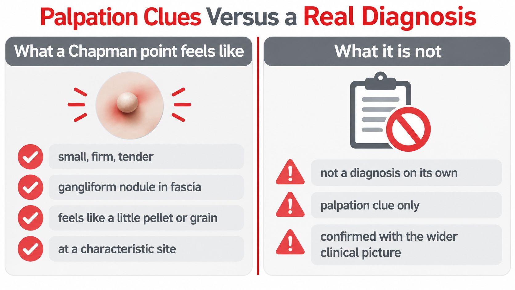
A Chapman point is traditionally described as a small, firm, tender, gangliform nodule in fascia, sometimes compared with a little pellet. That description helps students identify the intended examination concept. It doesn't make the finding specific for visceral disease.
The NIH-hosted clinical review of Chapman points reports minimal direct research and no definitive consensus or guideline regarding their function or clinical value. Its practical implication is straightforward: the finding should generate a hypothesis, not establish a diagnosis independently of history, vital signs, examination, and appropriate testing.
Separate similar-sounding findings
Chapman points compared with nearby exam findings
| Finding | Typical feel | COMLEX use | Does not establish |
|---|---|---|---|
| Chapman point | Small, firm, tender fascial nodule | Traditional organ pairing by location | Disease, cause, or treatment benefit |
| Counterstrain tender point | Focal tenderness in muscle or related tissue | A somatic dysfunction and a position of ease | A visceral diagnosis |
| Trigger point | Taut band with referred pain | Myofascial pain pattern | A Chapman organ map |
| Viscerosomatic segmental change | Tissue texture change, asymmetry, restriction, tenderness | A level that may relate to a viscus | One specific Chapman point |
These categories answer different examination questions. A counterstrain item may ask about positioning; a trigger-point item may emphasize referred pain; a Chapman item may ask for a traditional organ association. The word tender alone doesn't identify which framework applies.
Broader viscerosomatic findings also shouldn't be reduced to a Chapman chart. A segmental tissue change describes a different level of observation from a discrete nodule at a named site.
Keep the clinical hierarchy intact
A defensible differential diagnosis depends on the whole presentation. Students should apply these rules when an item mixes palpatory findings with clinical evidence:
- Prioritize specific evidence. Imaging, laboratory findings, instability, or a red flag shouldn't be overridden by a reflex point.
- Resolve anatomical ambiguity. When candidate organs share a neighborhood, the exact landmark and side may distinguish the taught association.
- Limit the inference. Association doesn't establish causation, diagnostic accuracy, or evidence that manipulation treats the associated organ.
A question about a traditional association and a question about the next clinical step may use the same finding but require different answers.
The chart isn't a method for diagnosing a person or deciding whether treatment should begin, stop, or change. Clinical assessment requires appropriately qualified care, particularly when symptoms are acute, progressive, or concerning.
Mnemonics and a Ten-Minute Retrieval Drill
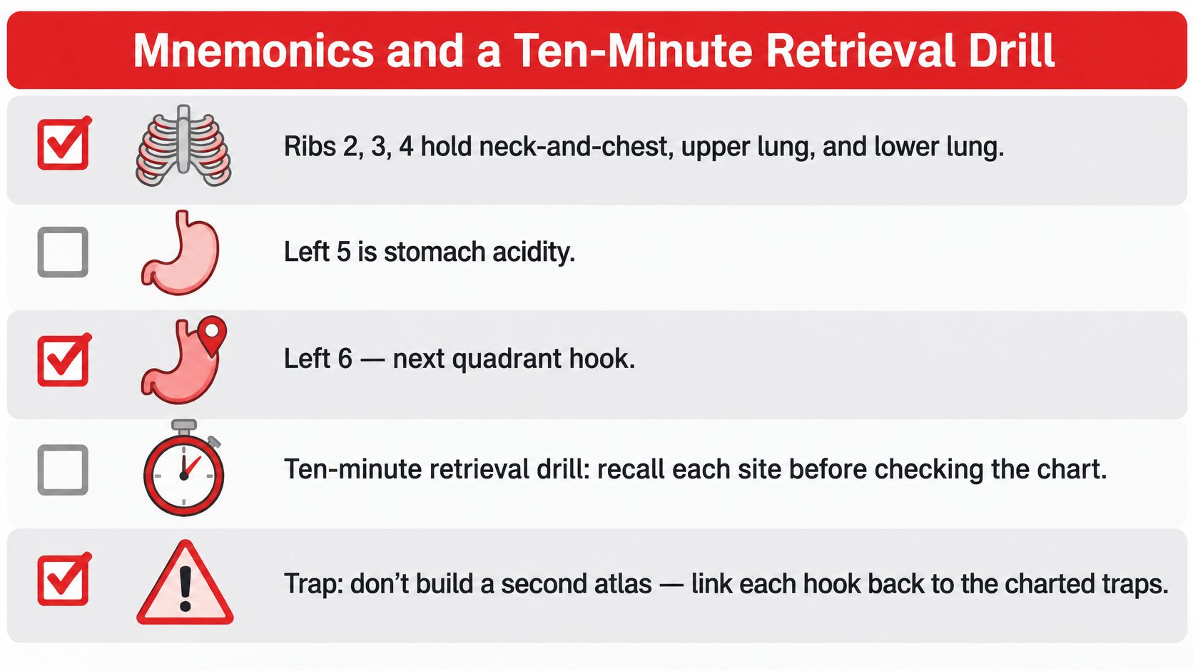
A useful mnemonic should preserve an anatomical distinction. A memorable phrase that drops the side or changes a rib into an interspace can make an error more persistent.
Students can organize recall around contrasts, rather than build a second atlas:
- Chest ladder. Group the upper chest associations separately from upper and lower lung.
- Left stomach pair. Store acidity and peristalsis together, but retain their different interspaces.
- Opposite-side pair. Contrast pancreas with spleen instead of learning each in isolation.
- Higher adrenal, lower kidney. Link relative anterior height with the corresponding posterior sequence.
- Appendix isn't cecum. Keep the rib-tip association separate from the proximal thigh association.
- Colon has different shapes. Anterior recall follows the iliotibial band; posterior recall uses a triangular region.
Produce an answer before checking it
The following ten-minute drill is a suggested study exercise, not a validated readiness measure:
- Prepare the page during the opening minute. Create columns for organ and both sites. Keep the charts out of view.
- Use minutes two through six for production. Write ten organs from memory, including anterior location, posterior location, and side.
- Check mismatches during minutes seven and eight. Compare only uncertain or incorrect entries with the course-approved atlas.
- Repair misses during minutes nine and ten. Rewrite each missed pairing once, then attempt it again without looking.
The next day's practice should begin with the same organs. Once those responses become reliable, another set can include pelvic points and previously confused neighbors.
This exercise exposes something highlighting doesn't: whether the student can generate the answer without seeing it. A student who recognizes “spleen” in a completed table may still struggle when a stem supplies only an interspace and side.
Reverse the direction
Organ-to-site recall and site-to-organ recall aren't identical tasks. Students should alternate them rather than assume that success in one direction guarantees success in the other.
For a missed response, the correction should name the exact distinction: wrong side, wrong interspace, wrong landmark, or wrong organ. Recopying an entire page after a single mismatch adds work without clarifying what caused the error.
Original Vignettes and Why an Answer Wins
The following are original, unofficial teaching examples. They aren't recalled, leaked, or official COMLEX questions, and no independent clinician review is claimed.
Each example separates the association being tested from what would be necessary to evaluate a real patient. People with abdominal pain, fever, or other red flags need prompt clinical evaluation; these examples aren't a diagnostic method.
Example A separates the appendix from the cecum
A student presents to student health with abdominal discomfort that began near the umbilicus and later localized to the right lower quadrant. The stem describes low-grade fever and a small tender nodule at the tip of the right twelfth rib.
Which traditional Chapman association is being tested?
The choices are appendix, cecum, ovary, and gallbladder.
Appendix is the intended answer. The decisive association is the named rib-tip landmark. The pain pattern supports the same organ, but the question asks for the traditional pairing rather than a definitive diagnosis.
The distractors reveal different errors:
- Cecum tests regional overgeneralization. Remembering that the cecum belongs in the right lower abdomen doesn't place its anterior Chapman association at the rib tip.
- Ovary tests organ proximity. A pelvic organ may enter a clinical differential, but that doesn't change the landmark named in this association question.
- Gallbladder tests right-sided anchoring. “Right side” is too broad; the gallbladder association belongs in a different anterior region.
A student who chose cecum needs a landmark correction, not necessarily a complete review of abdominal pathology. A student who diagnosed appendicitis solely from the nodule needs an interpretation correction.
Example B distinguishes adrenal from kidney
A patient reports fatigue and salt craving. An educational stem describes a tender nodule about two inches above and one inch lateral to the umbilicus, together with a posterior finding in the T11 region.
Which association fits better, adrenal or kidney?
Adrenal is the intended pairing. The anterior position is higher than the kidney teaching site, and the posterior description supports the adrenal region.
The symptoms don't establish a particular endocrine disorder. Fatigue is nonspecific, and the stem doesn't provide the information necessary for a complete diagnosis. The useful reasoning move is to answer the association question without expanding it into an unsupported disease conclusion.
For kidney, both anatomical details would need reconsideration: the closer anterior relationship to the umbilicus and the lower posterior teaching level. Missing both suggests that the student memorized the organ names without retaining their relative arrangement.
Example C checks a neighboring interspace
A short identification item describes a nodule at the left seventh intercostal space and offers stomach or spleen.
Spleen is the traditional pairing. “Left upper abdomen” isn't precise enough to make stomach the answer; the named interspace distinguishes the choices.
Review the error rather than the question's appearance
The NBOME scoring and reporting guidance states that scored questions contribute equally, apart from unscored pretest questions. An unfamiliar osteopathic manipulative medicine (OMM) stem therefore doesn't deserve extra time because it looks unusual.
A practical review log can classify misses as location, side, organ, or overinterpretation. Students using COMLEX Level 1 question-review guidance can apply the same classification to broader vignette practice.
Review priority: Correct the smallest identifiable reasoning error before adding more material to the study plan.
Build a Chapman Point Recall Plan
Seven-day Chapman point recall cycle
How can a recall plan make point associations easier to retrieve without turning them into diagnostic conclusions? Use brief practice alongside broader organ-system review, and keep a clear evidence limit attached to each task. This cycle is a study suggestion, not an official examination schedule.
| Day | Focus | Recall target |
|---|---|---|
| Day 1 | Anterior chest | Middle ear through lower lung on the anterior chart; retrieve the listed site and organ pairing |
| Day 2 | Anterior abdomen | Stomach through pancreas and spleen; check the side as well as the organ |
| Day 3 | Iliotibial and pelvic points | Cecum through descending colon, ovary, testis, and uterus; treat each as a traditional association |
| Day 4 | Posterior cervical and thoracic | C1 through T10 levels on the posterior chart; recall the taught level without inferring a diagnosis |
| Day 5 | Posterior lumbar and colon triangle | T11 through L2 levels, kidney, bladder, and colon triangle; separate location recall from clinical conclusions |
| Day 6 | Mixed retrieval | Recall ten organs from memory with both sites; use the chart to check errors afterward |
| Day 7 | Missed rows only | Rewrite only rows missed during the preceding days, including the specific location or side error |
Match review to the broader blueprint
The NBOME Level 1 specifications assign minimums of 12% to Osteopathic Principles, Practice, and Manipulative Treatment and 6% to Osteopathic Patient Care and Procedural Skills. These figures describe domain allocations, not the share devoted specifically to Chapman points.
That distinction guides study time. A domain percentage does not mean one location chart deserves a similar proportion of preparation. Place Chapman review alongside other osteopathic concepts, biomedical sciences, and clinical reasoning.
The NBOME examination-format page describes Level 1 as a one-day, eight-hour computer-based examination with 320 single-best-answer questions, divided into separately timed four-hour sessions. Unused time from the first session does not transfer to the second. Recheck the exam format and specifications on the NBOME site before scheduling.
Adjust the task by examination level
For Level 1 preparation, schedule a short anterior review block and a separate posterior-and-pelvic block within the weekly system review. Connect each location to anatomy and relevant pathology, then test recall without relying on list recitation.
For Level 2-CE preparation, focus on what the clinical question asks. A Chapman point may serve as a supporting clue, while diagnosis or management depends on the rest of the presentation.
For Level 3 preparation, use a maintenance pass to retain familiar pairings and leave more study time for clinical decisions. This is a planning recommendation, not a claim about how often Chapman questions appear at any level.
A recall block can become shorter when you can:
- Identify the landmark without confusing a rib, interspace, or transverse-process region.
- Supply the side without checking the chart.
- Name the competing association that previously caused a miss.
- State the evidence boundary without turning the point into a diagnosis.
For structured support, COMLEX tutoring information describes help with content review, clinical reasoning, question approach, and pacing for Level 1, Level 2-CE, and Level 3, including students facing a plateau or returning after an unsuccessful attempt.
A difficult study block or prior attempt does not determine a medical career.
Sources
- NCBI Bookshelf clinical review of Chapman points supports the historical description and discussion of the evidence limits. Use it to distinguish traditional associations from diagnostic conclusions.
- NBOME COMLEX-USA Level 1 specifications describe the osteopathic competency domains and integrated examination framework.
- NBOME COMLEX-USA examination format explains the Level 1 format and session timing.
- NBOME COMLEX-USA scoring and reporting explains how scored questions contribute to the reported result.
The charts are study aids. Check point conventions against your assigned OMM text and school syllabus, and treat a palpatory association as one clue rather than a diagnosis.
Ace Med Boards offers COMLEX tutoring in content review, clinical reasoning, question approach, and pacing for Level 1, Level 2-CE, and Level 3. Students can schedule a free COMLEX consultation to discuss their exam level and timeline.