Immunology is the study of how the body distinguishes self from non-self and mounts specific defenses, and the fastest way to make it exam-proof is to learn one reusable immune-response map. If you can redraw that map from barrier defenses to resolution or pathology, the rest of the subject starts to organize itself.
What Immunology Actually Tests on the Board Exams
Immunology on USMLE, COMLEX, and related exams is rarely a straight recall test. It's a pattern-recognition test built around a few recurring scenes, a child with recurrent infections, a transfusion reaction, a wheal-and-flare rash, a vaccine question, or a patient whose labs point to complement failure.
The board writer usually wants you to do four things at once. First, identify the cell type or surface marker involved. Second, link a mediator to the mechanism, such as antibody, T cell, cytokine, or complement. Third, predict what infection pattern or pathology follows from the defect. Fourth, translate the vignette clue into the correct immune compartment, not just the correct buzzword.
The single map that keeps all of that organized has six stations, barrier, innate recognition, antigen presentation, lymphocyte activation, effector output, and resolution or pathology. Once you know where a clue enters the map, the answer options get much easier to narrow. A good immunology question usually breaks because the reader misplaces the defect on the map, not because the subject is obscure.
For learners preparing with the official exam blueprint, it helps to keep the tested topics anchored to the broader content outline used for board study, including the immunology-related material on the USMLE content outline.
Practical rule: When a stem feels broad, ask, “Which station of the immune-response map is broken?” That question is usually more useful than memorizing another isolated fact.
The Immune-Response Map From Barriers to Adaptive Effectors
Barrier and innate recognition
The first station is the barrier layer. Skin, mucosa, low pH, lysozyme, defensins, and commensal competition all make it harder for microbes to establish themselves in the first place. If that layer fails, the next responders are innate cells and molecules, not antigen-specific lymphocytes.
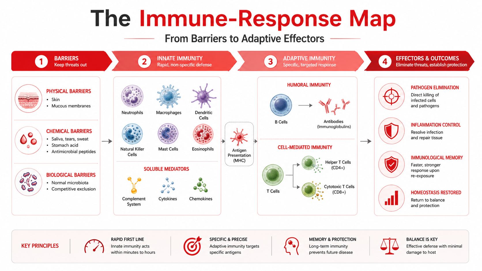
Innate immunity is fast, broad, and built to recognize patterns, not unique epitopes. Macrophages, neutrophils, dendritic cells, NK cells, and complement proteins detect danger through pattern-recognition logic, then begin inflammation, phagocytosis, or lysis. A useful external visual for comparison is the virusfaq.com immune diagram, which can help you place the moving parts before you drill the details.
Antigen presentation and lymphocyte activation
The handoff from innate to adaptive immunity happens when dendritic cells engulf antigen, mature, migrate to lymph nodes, and present peptide on MHC II to CD4 T cells. Intracellular peptides are presented on MHC I to CD8 T cells, which is why viral or tumor antigens so often point toward cytotoxic responses.
That activation step is also where cytokines begin to steer the type of adaptive response. IL-12 favors Th1, IL-4 favors Th2, and TGF-beta plus IL-6 favors Th17. Those choices matter because they determine whether the answer is cellular killing, antibody production, or mucosal/inflammatory recruitment.
High-yield anchor: If you can identify the antigen source, extracellular or intracellular, you can usually predict the MHC pathway and the dominant T-cell branch.
For a quick review of how immune concepts fit into microbiology-style organism clues, it also helps to revisit the linked microbiology review hub and connect microbes to host response rather than studying them in isolation.
Hypersensitivity and Immune-Complex Reasoning
| Type | Mediator | Timing | Mechanism | Classic Examples |
|---|---|---|---|---|
| I | IgE, mast cells | Immediate | Allergen cross-links IgE on mast cells, causing histamine release | Anaphylaxis, atopy, wheal-and-flare |
| II | IgG or IgM | Hours to days | Antibody binds a cell-surface or matrix antigen, causing complement, opsonization, or altered receptor function | Hemolytic transfusion, Goodpasture, myasthenia gravis, rheumatic fever |
| III | IgG or IgM immune complexes | Hours to days | Soluble antigen-antibody complexes deposit in tissues, activate complement, and recruit inflammation | Serum sickness, SLE, post-streptococcal glomerulonephritis, Arthus reaction |
| IV | T cells | Delayed | T-cell mediated inflammation, no antibody required | Contact dermatitis, tuberculin skin test, granulomas |
The easiest way to separate type II from type III is to ask three questions. Where is the antigen? If it sits on a cell surface or matrix, think type II. If it's soluble and circulating, think type III. Which effector causes injury? Antibody directed at a target cell suggests type II, while immune-complex deposition suggests type III. Where does the damage appear? Target-cell injury points to a specific tissue or blood cell, while immune complexes often show up in vessels, kidneys, or joints.
A hypothetical acute hemolytic transfusion reaction makes the distinction clean. A patient receives an incompatible unit, then develops fever, flank pain, hypotension, hemoglobinuria, and dark urine soon after transfusion, with intravascular hemolysis from antibody binding donor red-cell antigen and activating complement. That is type II hypersensitivity, not immune-complex disease, because the antibody is reacting against a cell-bound target rather than forming circulating complexes.
Exam habit: If the stem gives you a target cell, think type II. If it gives you a precipitating complex that deposits in tissue, think type III.
For official background on immune-mediated injury patterns, a useful mechanistic reference is the CDC and NIH-linked immunology material on antibody and tissue injury concepts, especially when you want the complement and inflammatory logic tied together.
The Complement Cascade in Five Rungs
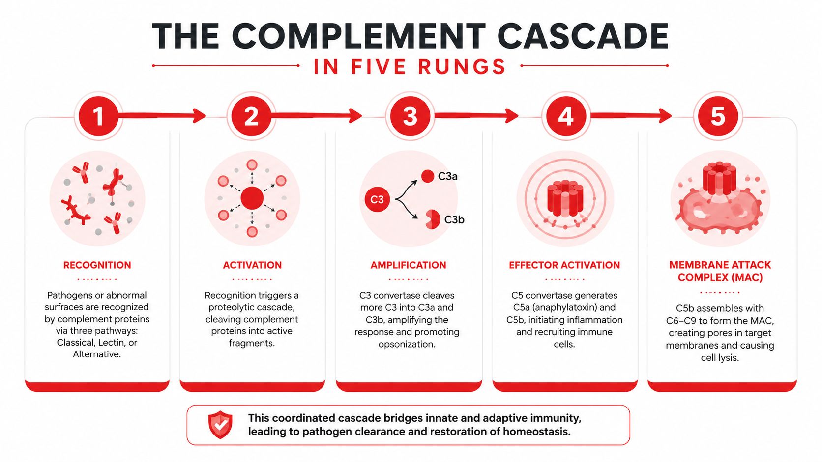
Complement works best as a ladder, not a paragraph. Rung 1 is initiation, Rung 2 is convertase assembly, Rung 3 is C3 splitting, Rung 4 is amplification and C5 cleavage, and Rung 5 is terminal membrane attack.
The three named pathways are classical, lectin, and alternative, and they all converge at C3 cleavage. Classical starts with immune complexes, lectin starts with carbohydrate recognition, and alternative runs as a surface-amplifying pathway. The convergence at C3 is the key amplification step, and downstream C5 cleavage drives the strongest inflammatory and terminal effects (NCBI Bookshelf complement review).
A compact way to redraw the cascade is this:
- Classical trigger: immune complexes bind C1q
- Lectin trigger: mannose-binding lectin recognizes microbial sugars
- Alternative trigger: spontaneous C3 tick-over on microbial surfaces
- C3 products: C3b opsonizes, C3a is anaphylatoxic
- C5 products: C5a is a strong inflammatory and chemotactic signal, C5b begins the terminal pathway
- Terminal effect: C5b-9 forms the membrane attack complex
The clinical clues attach to specific rungs. Hereditary angioedema reflects C1 inhibitor loss, so the classical pathway's early brake is missing. Paroxysmal nocturnal hemoglobinuria comes from absent CD55/CD59, which allows complement injury on blood cells. C1q deficiency points toward lupus-like autoimmunity because immune-complex handling is impaired.
The most testable infection patterns follow the end of the ladder. C3 failure usually means recurrent severe pyogenic infection, while terminal C5 through C9 deficiency points toward Neisseria susceptibility. That distinction is worth memorizing as a pathway problem, not as two disconnected facts.
For biochemical context on why this cascade amplifies so quickly, the biochemistry review page is a helpful companion when you want to connect proteins, cleavage, and downstream inflammation.
Immunodeficiency and Transplant Patterns
Primary immunodeficiencies become easier when you group them by where the immune map breaks. A barrier or neutrophil defect suggests trouble with the earliest antimicrobial work. A B-cell defect suggests poor humoral defense and weak antibody responses. A T-cell defect suggests broader vulnerability because cellular coordination fails too.
| Defect Branch | Key Condition | Hallmark Cue | Characteristic Organism or Finding |
|---|---|---|---|
| Phagocyte | Chronic granulomatous disease | Defective oxidative burst | Catalase-positive organisms |
| Phagocyte | Leukocyte adhesion deficiency | Delayed umbilical-cord separation | Poor neutrophil migration |
| Phagocyte | Chédiak-Higashi syndrome | Giant granules, partial albinism | Impaired lysosomal trafficking |
| B cell | Bruton agammaglobulinemia | After maternal IgG wanes | Recurrent bacterial infections |
| B cell | Common variable immunodeficiency | Poor vaccine response in adulthood | Hypogammaglobulinemia |
| B cell | IgA deficiency | Mucosal infection risk, transfusion reactions | Anaphylaxis with blood products |
| T cell | DiGeorge syndrome | 22q11 deletion, thymic hypoplasia | Low T cells, hypocalcemia |
| T and B cells | Severe combined immunodeficiency | Early severe infections | Profound combined defect |
The transplant timeline follows the same logic. Hyperacute rejection happens when preformed antibody attacks the graft quickly, often with thrombosis. Acute cellular rejection is driven by T cells. Acute humoral rejection involves donor-specific antibodies. Chronic rejection produces fibrosis and vascular intimal thickening over time. Once again, the question is less “what is the label?” and more “which branch of the immune-response map is overactive or absent?”
For a current overview of broader immunology trends and human-based model systems, the human-relevant models discussion is useful background, especially when you're thinking about why immune testing and transplant biology are increasingly tied to human tissue and translational design.
Vaccines, Adjuvants, and the Adaptive Memory Map
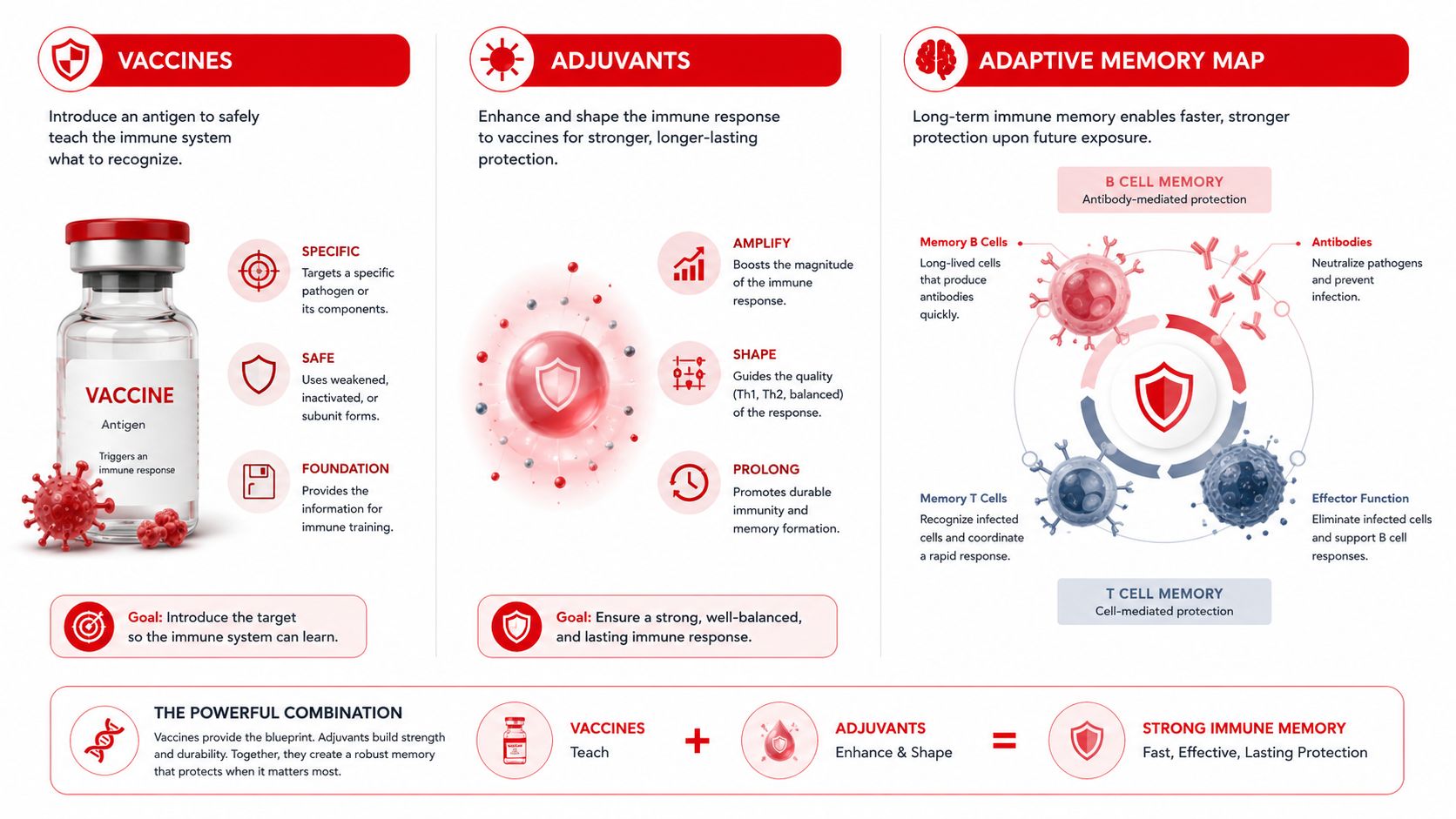
Vaccines are easiest to remember when you attach each platform to the station on the map it manipulates. Live attenuated vaccines such as MMR, varicella, yellow fever, and intranasal influenza replicate enough to create strong cellular and humoral memory, which is why they're avoided in pregnancy and severe immunodeficiency. Inactivated and toxoid vaccines such as Hep A, rabies, and inactivated polio are safer but usually depend more on boosters because they mainly generate antibody-based memory.
Subunit, conjugate, and recombinant vaccines lean heavily on adjuvants because purified antigen alone can be weakly immunogenic. Adjuvants work by amplifying the innate alarm, strengthening antigen uptake, improving co-stimulation, and helping the adaptive response cross the threshold for durable memory. Conjugate vaccines matter because they convert a T-independent polysaccharide into a T-dependent response, which is why they work better in young children.
A compact comparison helps when the stem mixes platforms:
- Live attenuated: strongest cellular plus antibody memory, but not for severely immunocompromised patients
- Inactivated or toxoid: safer, usually booster-dependent
- Subunit or recombinant: purified antigen, often adjuvant-dependent
- Conjugate: polysaccharide linked to protein to recruit T-cell help
- mRNA: antigen is translated in host cells, and the RNA itself can act as an innate signal through TLR7/8
Passive immunity is a different mechanism entirely. Maternal IgG and immunoglobulin products provide immediate protection, but they do not build memory because the recipient's own lymphocytes were not trained through active antigen exposure.
If you want a structured way to retain vaccine logic across spaced review, the spaced repetition guide pairs especially well with antigen-class questions and memory-based recall.
Worked Practice Vignettes for Immunology
A young child keeps getting infections with encapsulated bacteria. The key question is whether the defect sits in the antibody arm or the complement arm. If the stem also mentions poor vaccine responses and recurrent sinopulmonary infections, a B-cell problem becomes more likely; if it emphasizes complement susceptibility, think about the cascade and the organisms that reveal it.
A second stem uses a delayed rash after a tattoo or jewelry exposure. The clue is not the skin lesion alone, it's the timing and the trigger. That points to type IV hypersensitivity, with T cells as the effector cells and delayed inflammation as the mechanism.
A third stem involves maternal-fetal Rh incompatibility. The antibody that crosses the placenta is IgG, not IgM, and the prevention strategy works by blocking maternal sensitization before it can seed future hemolysis. The point is to connect antibody class with placental transfer, not to memorize the disorder in isolation.
A few original practice anchors can make the map stick:
- Encapsulated infection in a child. Ask whether the issue is poor opsonization, poor antibody production, or impaired phagocyte killing.
- Delayed contact reaction. Ask whether the mechanism is antibody-mediated or T-cell mediated.
- Maternal-fetal hemolysis. Ask which immunoglobulin crosses the placenta and why the first exposure matters less than the subsequent one.
The answer is usually in the timing, the antigen source, and the effector arm. Those three clues separate most immunology stems faster than trying to remember the disease name first.
If you want a more personalized way to turn misses into a durable recall system, Ace Med Boards also offers one-on-one immunology review as part of its broader board-prep tutoring, which can be useful when your misses cluster around hypersensitivity, complement, or vaccine reasoning rather than content volume alone.
High-Yield Study Plan and Question-Review Workflow
| Week | Focus Topics | Daily Block Structure | Weekly Milestone |
|---|---|---|---|
| 1 | Barrier defenses, innate recognition, cells, cytokines | Concept review, retrieval, 10 to 15 questions | Draw the full immune-response map from memory |
| 2 | Antigen presentation, MHC, T cells, B cells | Concept review, retrieval, 10 to 15 questions | Explain MHC I versus MHC II without notes |
| 3 | Hypersensitivity and complement | Concept review, retrieval, 10 to 15 questions | Sort type II versus type III on cue |
| 4 | Immunodeficiency, vaccines, transplant | Concept review, retrieval, 10 to 15 questions | Tie defect, organism, and rejection pattern together |
A daily 45-minute block works well when it's split into three parts. Spend the first segment on concept review, the second on active recall or spaced repetition, and the last on board-style questions. Use Saturday for a longer mixed block and Sunday for an error-log pass, because immunology errors usually repeat in the same few forms.
The question-review workflow should be mechanical. Tag every miss by map rung, mediator, or clinical keyword. Then convert those tags into next-day micro-cards so the same mistake becomes a retrieval cue instead of a repeated loss.
Here's the simplest scoring approach for your error log:
- Knowledge miss: You didn't know the fact, so add it to a micro-card.
- Reasoning miss: You knew the fact but placed it on the wrong map rung, so redraw the pathway.
- Misread miss: The stem clue was there, but you rushed past it, so rewrite the clue in plain language.
- Pacing miss: You reached the right idea too slowly, so practice faster pattern sorting.
For a broader step-wise plan that fits the rest of your board schedule, the USMLE Step 1 study plan gives a useful framework for where immunology fits alongside the other foundational sciences.
If you want guided help turning these immune patterns into a cleaner exam strategy, Ace Med Boards offers focused board tutoring and structured question analysis for medical students preparing for USMLE and COMLEX. If you'd like a personalized plan for your immunology weak spots, visit Ace Med Boards and schedule a free consultation.