You're staring at a long question stem, the lab values look busy, and three answer choices all sound plausible. That's exactly where pathophysiology matters most, because it helps you work backward from the clinical clue to the mechanism that made the patient look that way in the first place. For medical students, the fastest way to make this topic usable is to treat it as a cause-to-sign translation system, not a list of diseases to memorize.
Pathophysiology is the study of disordered function in disease, including the causal chain from the initial insult to the body's compensatory response and the final symptoms, signs, and lab changes. In practical board prep, that means asking a simple question every time: what changed in normal physiology, and why does that change produce this presentation? That framework matters because pathophysiology is really the bridge between physiology and clinical reasoning, the same bridge Hecker helped formalize in the late 18th century when the field began taking shape as its own discipline (historical overview).
What Pathophysiology Really Means for Med Students
Start with the difference between normal function and disease
Physiology tells you how a healthy system works. Pathology tells you what structural abnormality is present. Pathophysiology explains how the abnormality disrupts function, and that distinction is exactly where exam questions live. A coronary plaque is pathology, but the downstream ischemia, chest pain, and troponin rise are pathophysiology in motion.
That difference sounds small until you hit a board-style vignette. A 58-year-old with chest pain, diabetes, and diaphoresis does not need a vocabulary test. You need to decide whether the stem is pointing to ischemia, reflux, anxiety, or something else, then trace the mechanism from oxygen supply-demand mismatch to the findings in front of you. A useful way to think about it is this, pathology names the lesion, physiology names the normal system, and pathophysiology tells you why the system failed.
Practical rule: if the answer choice is a diagnosis, the mechanism is usually hidden in the stem.
Why boards reward mechanism more than recall
Board exams rarely ask for isolated facts in a vacuum. They ask whether you can connect a disruption in function to a predictable clinical pattern. That's why mechanism-first reasoning beats fact-list memorization, especially when two answer choices both sound familiar.
The history of the field reflects that same shift. Pathophysiology emerged as a distinct discipline in the late 18th century, with first lectures in 1790 at the University of Erfurt and the first known textbook in 1791, which ran to 770 pages (historical record). By the 1930s, the subject had become a formal part of medical school curricula, reflecting the move from naming disease to explaining disease mechanism (PubMed historical review).
For your own studying, that means every vignette should become a causal chain. First identify the normal pathway. Then ask what broke, what the body tried to do about it, and why those compensations created the symptoms you see. If you keep that pattern in mind, the rest of this guide becomes a toolkit instead of a wall of facts. For a related framework on clinical reasoning, see this Ace Med Boards guide to clinical reasoning.
The Five Core Mechanisms Behind Most Diseases
The recurring toolkit behind high-yield disease
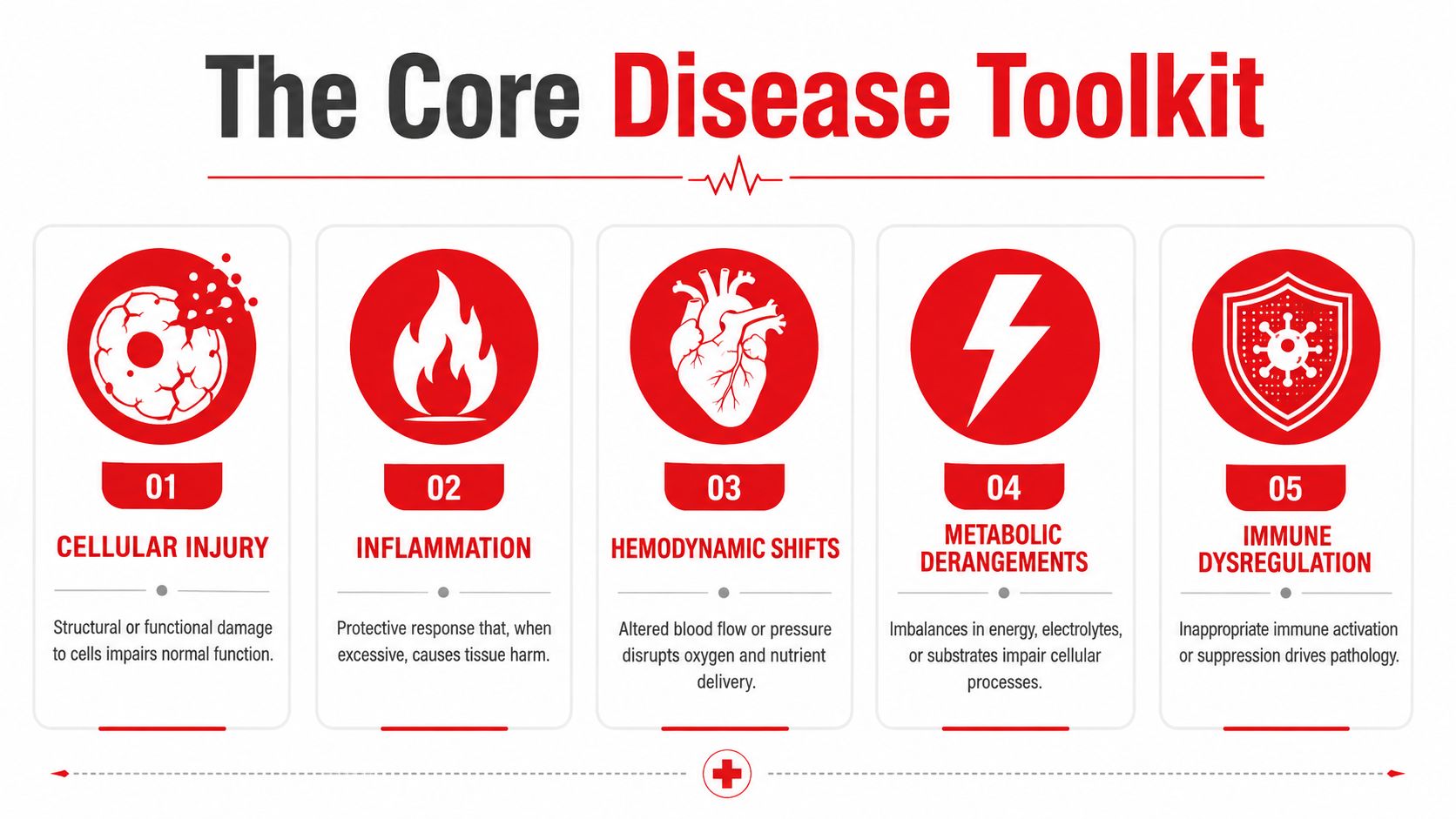
A lot of diseases look different on the surface but collapse into a small set of recurring mechanisms. Cellular injury, inflammation, hemodynamic shifts, metabolic derangements, and immune dysregulation explain a surprisingly large share of the disease patterns you'll see on exams. General teaching in pathophysiology emphasizes exactly that kind of shared-mechanism thinking, including hypoxia or ischemia, abnormal cell proliferation, inflammation, and metabolic abnormalities that create toxic or undernourished environments (core-process framework).
The point isn't to reduce every disease to one label. The point is to recognize the mechanism that dominates the stem. Is a membrane failing because of ATP depletion? Is the body overreacting with cytokines? Is blood flow inadequate? Is metabolism misfiring because insulin signaling is broken? Is the immune system attacking the wrong target? Those are the big buckets.
Five mechanisms, five familiar examples
Cellular injury shows up when ischemia cuts ATP production and the Na/K pump fails, which leads to early cell swelling, a classic early event in myocardial infarction. Inflammation is the body's coordinated response to danger signals, and the inflammatory cascade can amplify leukocyte activation, cytokine release, and tissue remodeling (inflammatory signaling review).
Hemodynamic shifts matter when the problem is not the cell itself but the flow around it. Reduced preload lowers stroke volume, and that is why hypovolemic shock changes perfusion so quickly. Metabolic derangements are obvious in type 1 diabetes, where insulin receptor failure or absent insulin signaling drives lipolysis and ketoacidosis. Immune dysregulation appears when autoantibodies attack acetylcholine receptors in myasthenia gravis, weakening neuromuscular transmission.
Acute inflammation follows a reproducible sequence, fluid exudation, neutrophil infiltration, vasodilatation, pain, and loss of function, while mediator networks can keep injury going unless anti-inflammatory feedback restores homeostasis (acute inflammation review). That homeostatic-imbalance idea is the exam target. Disease often means a regulatory system has tipped beyond its compensatory range, and the symptom pattern is the body's failed attempt to recover equilibrium.
The board question usually isn't asking, “What disease is this?” It's asking, “Where did homeostasis break?”
Normal Versus Disease at the Mechanism Level
See the same system before and after failure
A table helps because a lot of students know the disease but not the normal pathway that precedes it. If you can describe the healthy mechanism first, the diseased version becomes much easier to reason through. That's especially true for cardiovascular, endocrine, and acid-base questions.
| Normal physiology | Disease mechanism |
|---|---|
| Cardiac output regulation: sympathetic tone raises heart rate and contractility when demand increases. | Heart failure with reduced ejection fraction: the ventricle cannot maintain forward flow, so RAAS activation and sympathetic compensation increase preload and afterload, which can worsen congestion. |
| Glucose homeostasis: insulin promotes glucose uptake through GLUT4 translocation and supports storage over breakdown. | Type 1 diabetes: absolute insulin deficiency removes that brake, so lipolysis increases and ketone production can drive diabetic ketoacidosis. |
| Acid-base balance: the kidney reclaims bicarbonate and secretes acid to preserve pH. | Metabolic acidosis: bicarbonate is consumed or lost, anions accumulate, and compensation cannot fully normalize the internal environment. |
The key lesson is that the disease column is not a different system. It is the same system pushed past its compensatory range. Once you start thinking that way, the stem gets less mysterious. A question about RAAS is rarely just about hormones, for example, it's usually about a heart or kidney system trying and failing to preserve perfusion.
Where students often miss the pivot point
The pivot point is usually the step where regulation stops working. In glucose physiology, the break is at insulin availability or response. In acid-base problems, it's often at buffering, renal handling, or respiratory compensation. In heart failure, it's the mismatch between output and the body's attempt to preserve blood pressure and perfusion.
That same logic is why the body's response can become part of the problem. Compensations are helpful in the short term, but they can create fluid retention, vasoconstriction, or metabolic stress when they persist. If you keep asking “what is the body trying to protect?” you'll start seeing the disease as a failed adaptation, not just a diagnosis.
Walking a Composite Clinical Vignette From Symptom to Mechanism
A composite case that makes the chain visible
Composite clinical vignette. A 58-year-old man presents with progressive dyspnea, orthopnea, bilateral lower-extremity edema, and an S3 gallop. The symptom pattern points to impaired forward flow and fluid backup, not a random collection of complaints. Once the ventricle weakens, the body responds by activating sympathetic signaling and RAAS, which raises preload and afterload in an effort to preserve circulation.
That compensation makes sense at first, but a failing left ventricle cannot handle the added volume and resistance. Pressure backs up into the pulmonary circulation, pulmonary capillary wedge pressure rises, and fluid begins to move into the lungs. That is why the patient feels short of breath, especially when lying flat, and why orthopnea and dyspnea fit the same mechanism rather than separate diagnoses.
The edema follows the same logic. When venous pressures rise and fluid handling changes, interstitial spaces become the place where excess volume shows up. An S3 gallop also fits the story because it reflects abnormal ventricular filling in a volume-overloaded heart.
For a similar case-based approach in study review, see case-based learning in medicine.
Why the same template shows up in other diseases
The same fluid-shift logic shows up outside cardiology. In cirrhosis, ascites reflects altered forces that push fluid into the peritoneal cavity. In nephrotic syndrome, edema also reflects disrupted balance between vascular and interstitial compartments. The exact trigger differs, but the exam logic stays the same, identify where the pressure, protein, or volume relationship has gone wrong, then trace the visible finding back to that break.
Clinical shorthand: symptom clusters matter because they reveal the failed compensation, not just the final diagnosis.
A Step 1 stem often hides this mechanism in plain sight. If the question gives dyspnea, orthopnea, edema, and an S3, you do not need to hunt for an exotic explanation. You need to recognize a heart that can't empty effectively and a body that is trying, unsuccessfully, to preserve circulation by retaining fluid and tightening vessels.
Translating Mechanism Into Board-Question Clues
A three-question checklist for every stem
When you read a vignette, ask three questions in order. First, which core mechanism is dominant. Second, what compensatory response is the question testing. Third, where has homeostasis tipped past its set point. Those three prompts keep you from chasing distraction clues.
Clues often arrive as effects rather than causes. Dyspnea plus edema plus hepatomegaly points toward right-sided congestion. Kussmaul respirations are a physical clue that the body is trying to compensate for metabolic acidosis. A lab abnormality such as a high anion gap points you toward a specific acid-base problem rather than a vague “sick patient” interpretation.
A quick clue map
- Symptom cluster: dyspnea, edema, and abdominal fullness usually suggest a shared hemodynamic problem.
- Physical finding: crackles, JVD, or an S3 can reflect pressure or volume overload rather than a primary lung disorder.
- Laboratory anchor: anion gap, BNP, or ferritin can point to the mechanism that drives the presentation.
- Response clue: tachycardia, tachypnea, or RAAS activation often signals compensation, not the root cause.
If you see a question that mentions edema, don't stop at “fluid overload.” Ask whether the cause is Starling-force imbalance, lymphatic obstruction, kidney loss of protein, or a neurohormonal response to low effective perfusion. That one habit can flip a wrong answer into a right one.
For a tighter way to sort out why distractors look tempting, review this distractor-analysis guide.
Common Exam Pitfalls and Persistent Misconceptions
Why good students still miss these questions
Medical education research shows that misconceptions can persist even in senior students across respiratory, cardiovascular, and homeostatic topics, which means ordinary review isn't always enough to fix them (misconception review). That's why a student can know the definition of a disease and still miss the question when the stem is reworded. The problem is often not knowledge loss, it's a fragile mental model.
One well-known trap is Frank-Starling confusion. Students may remember the curve but misplace the direction of the change, then choose the wrong explanation for heart failure or inotrope questions. Another trap is mixing up preload and afterload, which leads to errors when the question asks what changed first and what changed second. The fix is to anchor each term to a physical concept, filling versus resistance.
Wrong answer logic versus correct mechanism logic
| Common mistake | Correct framing |
|---|---|
| Edema means every patient is simply volume overloaded. | Edema can reflect Starling forces, lymphatic obstruction, renal protein loss, or neurohormonal retention. |
| Fever automatically means infection. | Fever can also reflect inflammatory signaling without a bacterial cause. |
| Shortness of breath always means the lungs are primary. | Dyspnea can be the downstream result of cardiac congestion, metabolic acidosis, or anemia. |
The misconception that matters most is treating symptoms as diagnoses instead of outputs. A body in trouble will often generate similar looking end points, like tachycardia, edema, or dyspnea, even when the initiating lesion differs. If your study method doesn't force you to trace the chain backward, the wrong answer choices start to look just as plausible as the right one.
For a helpful example of how compensation and clinical findings connect, ischemic heart disease literature explicitly shows inflammation as a response to myocardial ischemia, not an isolated event (ischemic heart disease review). That same causal logic is what the exam wants from you.
Building a Pathophysiology Study Plan That Sticks
Use a loop, not a one-time read
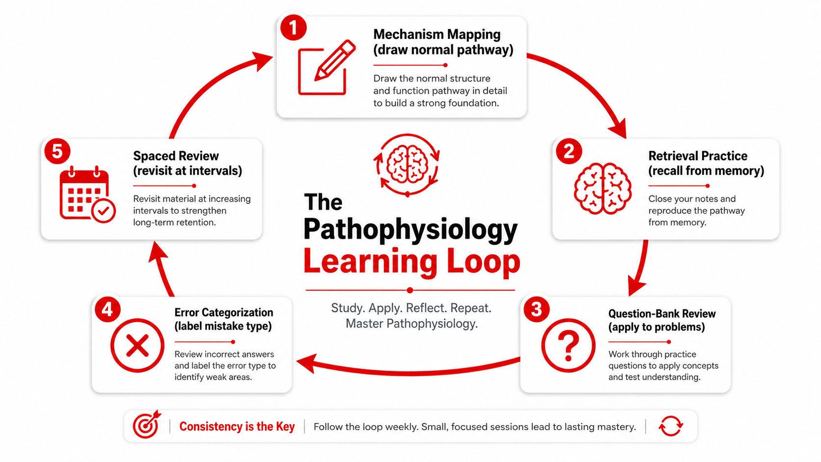
A durable study plan starts with the normal pathway, not the disease name. Draw the homeostatic pathway on a blank page, label the direction of flow, then mark where compensation should occur. After that, add one disease state, trace the lesion downstream, and write the symptoms, lab changes, and treatment logic in one chain.
A practical weekly workflow looks like this:
- Mechanism mapping: redraw the normal physiology from memory.
- Retrieval practice: explain the disease mechanism without notes.
- Question-bank review: connect the stem to the mechanism.
- Error categorization: label the miss as clue, mechanism, or answer-choice error.
- Spaced review: revisit the same concept later with a contrasting case.
The value of that loop is simple. You stop rereading the same explanation and start testing whether you can reconstruct it from a different angle. For spaced reinforcement, see this study guide on spaced repetition.
A simple weekly rhythm that keeps mechanism active
- Daily retrieval: spend a few minutes explaining one pathway out loud.
- Short question block: do a small set of stems and focus on mechanism, not just the answer.
- Error log review: sort every miss into the same three categories so patterns become visible.
- Weekly cumulative vignette: mix systems so you practice prioritizing clues under pressure.
If one topic keeps breaking, don't keep guessing at it in isolation. Rework the normal physiology first, then compare it with a diseased state and explain the shift in plain language. If needed, use a focused one-on-one session or a study partner to debug the specific point where your reasoning breaks.
Ace Med Boards offers diagnostic tutoring and personalized study-plan support that can fit this kind of mechanism-first review, especially when you need help turning missed stems into a cleaner error log. Visit Ace Med Boards if you want a low-pressure way to map out the next concept that needs clarification and build a pathophysiology plan around it.