Pathophysiology is the study of how disrupted normal physiology produces disease signs, symptoms, and lab changes, and it's the reasoning engine behind every USMLE and COMLEX vignette. If you can trace the mechanism, you can usually predict the next finding.
You've probably faced the same scene in a question bank or small group review, a tired-looking student, a long stem, and a multi-system problem that seems to reward memorization. The stronger move is to stop thinking of pathophysiology as a list and start treating it as a causal model, one that turns normal function into abnormal findings step by step.
What Pathophysiology Really Means on the Boards
A patient has fatigue, an unexpected laboratory abnormality, and a complication developing in the same organ system. A board-style question rarely asks whether you remember a definition. It asks you to trace the abnormality backward: which disrupted process explains this symptom, that lab change, and the next complication? That reasoning helps medical students, osteopathic medical students, international medical graduates, and residents move from mechanism to answer instead of from buzzword to guess.
Pathophysiology examines how normal biologic processes become abnormal and produce disease. Its historical roots include 1790, when Augustus Hecker gave the first lectures on the subject at the University of Erfurt, and 1791, when he published Grundriss der Physiologia pathologica, a 770-page work that reflected the field's development into a structured academic discipline rather than a loose overlap of pathology and physiology (historical overview).
A working definition you can use on exam day
Use this sentence: pathophysiology explains how an initiating abnormality produces disease through predictable changes in cells, tissues, organs, symptoms, and labs. It gives you a repeatable mechanism map when a question stem includes distracting details.
Write the chain in order:
- Normal function is disrupted.
- The body responds.
- That response changes tissue or organ function.
- The change produces clinical findings.
Inflammation may injure tissue, hypoxia may result from impaired oxygen delivery or impaired mitochondrial use, and type 2 diabetes may involve both insulin resistance and beta-cell dysfunction. These are connected mechanisms, not isolated facts (inflammation review, hypoxia review, type 2 diabetes review).
Practical rule: if you cannot explain the mechanism in one or two plain sentences, you probably do not own the concept yet.
Use Ace Med Boards' USMLE content outline when building a topic map. Organize each topic around the initiating defect, downstream response, and predicted findings.
On exam day, ask one question: what normal process was disrupted, and what should that disruption predict?
From Normal Physiology to Abnormal Findings
A patient becomes short of breath after losing blood. Another has the same symptom because alveoli cannot exchange gas. Both patients may show low oxygen levels, yet the disrupted physiology differs. Start with the normal process, then identify the point where the process fails.
That sequence turns a stem into a causal model. For perfusion, ventilation, filtration, signaling, or feedback, name the baseline first and the deviation second. Small shifts then become predictable findings rather than disconnected clues. A capillary that filters excess fluid produces a different pattern from one affected by lymphatic obstruction. Impaired gas exchange points toward a different mechanism than reduced oxygen delivery from low hemoglobin or low cardiac output.
Normal versus abnormal patterns
| System | Normal Physiology | Typical Pathologic Deviation | Resulting Clinical Clue |
|---|---|---|---|
| Capillary fluid balance | Fluid movement is kept in balance by opposing forces and drainage | Hydrostatic pressure rises, oncotic pressure falls, permeability increases, or lymphatic drainage is impaired | Edema, heaviness, swelling, site-specific accumulation |
| Ventilation and perfusion | Airflow and blood flow are matched for efficient gas exchange | Mismatch develops from airway, alveolar, or vascular disease | Hypoxemia, dyspnea, abnormal imaging or blood gas pattern |
| Acid-base balance | Lungs and kidneys keep pH in a narrow physiologic range | Acid production, CO2 retention, or renal compensation fails | Abnormal pH, compensatory respiratory or metabolic changes |
Use each row as a prediction tool. Ask three questions: What normally moves, matches, or compensates? Which variable changed? What finding should follow? This comparison habit turns a stem into a cause-and-effect puzzle and rewards reasoning over isolated recall.
Edema provides a useful worked example. Under normal conditions, the capillary acts like a controlled filter. Hydrostatic pressure pushes fluid outward, oncotic pressure draws it inward, permeability regulates passage, and lymphatic drainage removes the excess. If one force shifts, the location and character of the swelling can point toward a cardiac, renal, inflammatory, or lymphatic cause. A cardiovascular physiology guide can help reinforce the normal pressure relationships before you apply them to disease.
Now test the model with a counterfactual: if lymphatic drainage were restored while hydrostatic pressure remained high, would the edema disappear completely? No. It should improve, but the persistent pressure imbalance would continue pushing fluid into the tissue. Counterfactuals expose which mechanism is primary and which findings are downstream.
Study move: say the normal state out loud before naming the disease. Then complete the chain: disrupted process, physiologic response, organ-level change, clinical finding.
On exam day, ask: what normal process was disrupted, and what should that disruption predict? Use the Ace Med Boards' USMLE content outline to organize topic maps around those cause-and-effect steps.
The Major Mechanisms Behind Disease
Most board-relevant diseases can be organized around a few shared mechanisms. The language changes by organ, but the logic is familiar, inflammation, ischemia and reperfusion, cell injury and death, immune dysregulation, and genetic or metabolic causes. When you learn those mechanisms well, you stop relearning every disease from scratch.
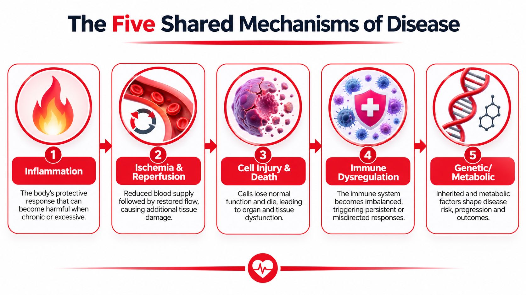
The cleanest teaching example is inflammation. It follows a concrete sequence, pattern receptors detect a harmful stimulus, inflammatory pathways turn on, mediators are released, and inflammatory cells get recruited (inflammation framework). That sequence matters because it gives you a repeatable way to reason through conditions that look different on the surface but share the same logic underneath.
Why inflammation keeps showing up
Inflammation is not just redness and swelling. Coordinated innate and adaptive immune signaling can drive tissue injury, oxidative stress, extracellular matrix remodeling, angiogenesis, and fibrosis across many organs (chronic inflammation review). In plain English, the host response itself can become part of the disease.
That's why many treatments are aimed at mediators rather than only the trigger. If the inflammatory cascade is doing the damage, then understanding the cascade helps you understand why suppression of a specific pathway can matter more than just naming the initial insult.
Why ischemia kills cells
Ischemia is another place where memorization fails fast. A major mechanism is calcium dysregulation, where increased cytoplasmic Ca2+ leads to cell injury, and lethal damage is driven largely by Ca2+-induced mitochondrial permeability transition, which depends on mitochondrial calcium accumulation (ischemic injury review). That is the reason ischemic cells die, not just a vague loss of oxygen.
Tissue hypoxia can also come from different routes. It may be hypoxic, anemic, or circulatory, all of which lower oxygen delivery, or it may be cytopathic hypoxia, where mitochondrial respiration is impaired even if global oxygen delivery looks preserved (hypoxia review). That distinction matters because fixing saturation, hemoglobin, or cardiac output won't necessarily solve cellular energy failure if the mitochondria are the problem.
The field keeps expanding into newer molecular subtypes too. Recent reviews discuss mechanisms such as ferroptosis, cuproptosis, nanodomain cAMP signaling, and redox signaling, but these are emerging explanatory models, not standalone memorization targets. Ace Med Boards' biochemistry resource fits well if you're trying to connect those mechanisms back to metabolism and cellular energy.
Mapping Mechanisms to Organ Systems
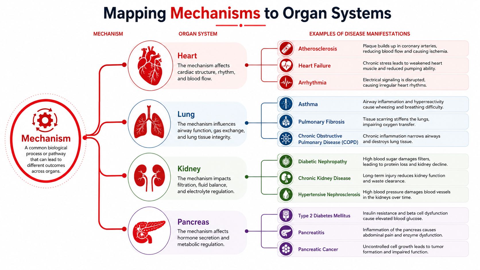
The same mechanism can look very different depending on the organ. That's why a smart student doesn't just learn “inflammation” or “insulin resistance,” but asks how that process behaves in the heart, lung, kidney, or pancreas. The organ gives the mechanism its clinical shape.
Edema as a teaching example
Edema is a good place to start because it forces you to think in forces, not labels. In normal physiology, the capillary is a controlled filter. If hydrostatic pressure rises, fluid gets pushed out. If oncotic pressure falls, fluid stays in the tissue space. If permeability increases, protein-rich fluid leaks out. If lymphatic drainage fails, the tissue can't clear what accumulates.
That gives you a practical way to sort findings. Cardiogenic edema points you toward increased hydrostatic pressure. Renal or protein-losing states make low oncotic pressure more likely. Inflammatory swelling suggests permeability changes. Localized swelling after surgery or nodal disruption points toward lymphatic problems.
Mechanism clue: edema is not one disease. It's a final common pathway, so the site, symmetry, and associated findings matter more than the word itself.
Diabetes and the logic of dual defects
Type 2 diabetes is another useful map because it's not a one-part problem. The literature describes two primary defects, impaired insulin secretion by pancreatic beta cells and reduced responsiveness of insulin-sensitive tissues to insulin (type 2 diabetes pathophysiology). Early on, beta cells can compensate by increasing insulin output, but hyperglycemia appears when they can't keep up anymore.
A more detailed review also links type 2 diabetes to gut microbiota, low-grade inflammation, lipid toxicity, mitochondrial dysfunction, beta-cell heterogeneity, incretin biology, and circadian rhythms, which helps explain why patients with the same diagnosis can behave differently (2025 diabetes review). Ace Med Boards' endocrine system guide is a reasonable companion if you want the endocrine framework organized by mechanism rather than by memorization buckets.
The same mapping habit applies to heart failure phenotypes too. Modern cardiovascular reviews separate HFpEF, HFmrEF, and HFrEF as distinct phenotypes shaped by inflammation, fibrosis, and neurohormonal imbalance (2025 cardiovascular review). The point isn't just naming the subtype, it's understanding why the same broad diagnosis produces different exam patterns.
Three Misconceptions That Stall Board Scores
A student faces a vignette with several familiar clues, yet cannot predict the next laboratory finding. The problem is often the mental model used to study Pathophysiology, rather than a lack of effort. Treat each disease as a causal model: identify the trigger, trace the mechanism, and predict the consequence.

Misconception one, Pathophysiology is a list
Isolated facts help you recognize terminology, but they do little when a question asks for the next finding. Build a mechanism map from normal physiology to the final clinical clue. Pair that map with active recall practice, which requires you to retrieve the explanation before checking the answer.
Misconception two, every disease is linear
Disease mechanisms branch and interact. Inflammation may promote fibrosis, alter neurohormonal signaling, and drive tissue remodeling, so one diagnosis can produce different patterns in different patients. A linear story can be a useful starting point, though it tends to break down once feedback loops join the picture.
Use a branching map: mark the initiating insult, the first affected process, the feedback loop, and the resulting organ findings. This keeps the mechanism flexible without turning it into an unstructured list.
Misconception three, buzzword recognition equals understanding
A classic phrase in a stem matters only when you can connect it to a mechanism. State the chain aloud: “This clue indicates this mechanism, which should produce this sign or laboratory pattern.” That sentence converts recognition into prediction.
A composite learner I often see in tutoring knows the disease list cold but freezes when asked what should appear next. More passive reading rarely solves that gap. Use prediction-before-answer drills, brief mechanism maps, and counterfactual questions such as, “If the beta cells were intact, what would change?”
Replacement habit: For every disease, write one sentence linking the trigger, mechanism, and expected clue. Then test the sentence by changing one condition and predicting which finding should move.
A Repeatable Mechanism Mapping Checklist
A patient has edema, fatigue, or an abnormal laboratory value. Rather than matching that clue to a memorized disease list, rebuild the causal model in the same order each time. The stable sequence keeps you asking what changed, how it changed, and what the change should produce.
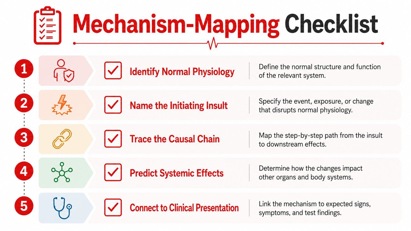
Use this sequence for any disease
- Identify normal physiology. State what the healthy organ, cell, or pathway normally does.
- Name the initiating insult. Decide whether the trigger is ischemia, immune activation, metabolic failure, obstruction, or another disturbance.
- Trace the causal chain. Follow the effect through cells, organs, and feedback loops.
- Predict systemic effects. Ask which symptoms, signs, and laboratory findings should follow.
- Connect to the clinical presentation. Match that mechanism to the pattern described in the vignette.
Consistency matters more than speed. The goal is a model you can reuse when the stem changes.
Worked example, edema
A stem gives you leg swelling and orthopnea. Begin with normal fluid balance, then test four possibilities: altered hydrostatic pressure, reduced oncotic pressure, increased permeability, or impaired lymphatic drainage. Orthopnea supports a cardiogenic pathway because increased filling pressures raise hydrostatic force and move fluid into tissues. Heavy protein loss or nephrotic-range clues shift the model toward low oncotic pressure.
Worked example, hyperglycemia
For hyperglycemia, separate insulin resistance from pancreatic compensation. A patient may remain near normal glucose while beta cells increase insulin output to meet demand. Rising glucose despite that response suggests beta-cell failure, because insulin secretion no longer matches the metabolic demand.
Apply the map after every question. Record the broken link: normal physiology, initiating trigger, causal mechanism, or clinical prediction. Then change one condition and predict which finding should change. This counterfactual test turns pathophysiology into causal reasoning rather than recognition alone.
Building a Pathophysiology Study Plan That Sticks
A durable plan isn't about rereading longer. It's about forcing retrieval, correcting weak links, and checking whether you can still reason when the stem changes. That matters for USMLE Step 1, COMLEX Level 1, and shelf prep, because board questions rarely reward familiarity alone.
Start by building a personal mechanism atlas. Pick one organ system at a time and write the normal state, the common insults, the causal chain, and the expected findings. Then use spaced repetition to revisit the same material after a delay, not immediately, so you can tell what stuck.
A simple question review workflow
- Name the failed step. Was the error in normal physiology, the insult, the chain, or the finding?
- Rewrite the mechanism in plain language. One or two sentences only.
- Add one counterfactual. Ask what would change if one variable were different.
- Test again later. Use delayed recall with a fresh vignette, not the same question.
That kind of review is more useful than just highlighting the correct choice. It shows you whether the concept is becoming automatic or whether it only feels familiar in the moment.
If you're also thinking about how medical learning content gets organized online, this Sight AI piece on content marketing in healthcare is a good example of how structure changes comprehension. The same principle applies to studying, when information is arranged around a clear framework, recall becomes easier.
A practical weekly rhythm
- Early week: learn one mechanism map from a trusted source.
- Midweek: close the notes and redraw it from memory.
- Later in the week: answer new questions that use the same mechanism in a different organ system.
- Weekend: review misses and identify the exact causal link you lost.
When board scores plateau despite solid content review, one-on-one tutoring can help by pressing you to explain the mechanism out loud, predict findings, and defend your reasoning under questioning. Ace Med Boards offers that kind of targeted support for students who want a structured second look at their pathophysiology reasoning, especially when content review alone isn't changing the way they answer questions.
If you want a clearer way to turn disease mechanisms into exam-day reasoning, schedule a conversation with Ace Med Boards and bring one topic that keeps slipping. A focused review of your mechanism maps can show you whether the issue is knowledge, recall, or the way you're connecting clues under time pressure.