
Isoelectric focusing separates proteins by isoelectric point, or pI, rather than mass. Each protein stops where the local pH equals its pI because its net charge is zero.
If a positive protein moves toward the negative electrode, why doesn't it finish at that electrode? That question exposes the key distinction in isoelectric focusing MCAT problems: initial movement and final band position aren't the same thing.
By Ace Med Boards Editorial Team
What Isoelectric Focusing Means on the MCAT
A premed reading a protein-separation passage may recognize the electrodes but still hesitate over the bands. The missing piece is often that a protein's charge changes with its surroundings. A protein doesn't carry one fixed net charge throughout an isoelectric focusing experiment.
Isoelectric focusing (IEF) separates proteins and other amphoteric molecules, which can act as acids or bases. Isoelectric point (pI) means the pH at which the molecule's positive and negative charges balance. Zero net charge doesn't mean every chemical group is uncharged.
A parking spot rather than an electrode finish line
A useful analogy is an assigned parking spot. The electrode determines the protein's initial direction of travel, but the protein's pI identifies its parking spot within the gradient.
Unlike a driver following a fixed route, the protein responds to its local chemical environment. As that environment changes, its protonation state and net charge change too. Its final location therefore reflects charge balance, not just the electrode it initially approached.
For Medical College Admission Test (MCAT) preparation, this connects amino-acid chemistry with experimental interpretation. Understanding protonation, pH, and the acid dissociation measure pKa makes a lab diagram easier to interpret without memorizing a laboratory protocol.
Key Takeaways
- Separation property: IEF sorts proteins by pI, not molecular mass.
- Below pI: A protein is net positive and moves toward the cathode.
- Above pI: A protein is net negative and moves toward the anode.
- Final position: The focused band is an internal equilibrium position, not a finish line at an electrode.
Every sample item in this article is an original teaching example, not an official Association of American Medical Colleges (AAMC) question. The useful reasoning habit is to identify the protein's present environment before predicting its movement.
The pH and pI Charge Rule
A protein's charge depends on how the pH at its current location compares with its pI:
| Local pH versus pI | Net charge | Moves toward | At equilibrium? |
|---|---|---|---|
| pH below pI | Positive | Cathode, the negative electrode | No, it keeps migrating |
| pH equal to pI | Zero | Neither electrode | Yes, it focuses here |
| pH above pI | Negative | Anode, the positive electrode | No, it keeps migrating |
The quick rule is: below pI, positive and toward the cathode; above pI, negative and toward the anode; at pI, no net migration. The MCAT.tools explanation of protein charge and electrode direction describes this relationship. In the standard electrophoresis setup, the cathode is negative and the anode is positive.
Why lower pH makes the protein more positive
Acidic surroundings favor protonation: ionizable groups bind protons. Some groups become positively charged, while negatively charged groups can become neutral. Together, these changes shift the protein's overall charge in the positive direction.
Basic surroundings favor deprotonation. Positively charged groups can lose their charge, while other groups become negatively charged. The protein's net charge shifts in the negative direction.
The rule describes the sum of charges across the protein. Individual side chains can behave differently, and a polar group is not automatically charged. The guide to polar amino acids helps distinguish polarity from charge.
Following a hypothetical protein through different environments
Consider a teaching example: a protein with a pI of 6.5. Its charge and initial direction depend on the pH where it is placed:
- At pH 4: The pH is below the pI, so the protein is net positive and initially moves toward the cathode.
- At pH 6.5: The pH equals the pI, so positive and negative charges balance. The protein has no net electrophoretic migration.
- At pH 9: The pH is above the pI, so the protein is net negative and initially moves toward the anode.
During focusing, those initial directions can change as the protein crosses the gradient. The final band marks the location where the local pH equals the pI, not the electrode the protein first approached.
Compare the pI with the pH at the protein's current location. The same pH could make one protein positive and another negative if their pI values differ.
Electrode names can cause a separate mix-up. For this electrophoresis setup, label the cathode negative and the anode positive before predicting movement.
For readers exploring earlier biology study options, funding an Access to Biology diploma is a separate educational-planning topic.
How a Protein Focuses Step by Step
A passage may describe ampholytes, sample loading, and an applied voltage in unfamiliar language. Those details become easier to follow when each event has a chemical reason.
The run in experimental order
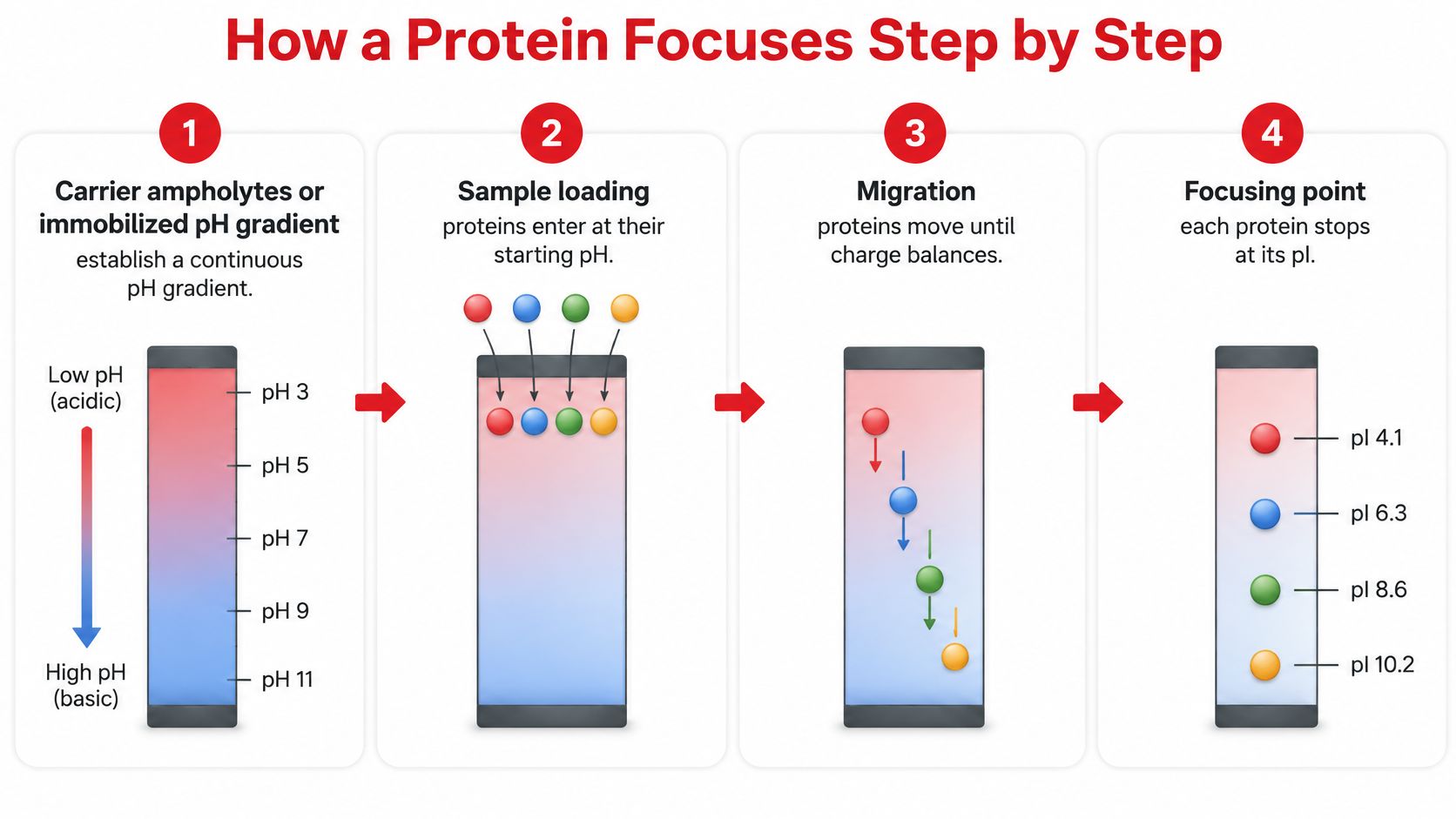
Establish the pH gradient. Carrier ampholytes can establish a continuous gradient under an applied field; an immobilized pH gradient provides another format. The Stanford microfluidics IEF overview describes experimentally selected gradients such as pH 1–12, rather than a universal range required for every experiment.
Place the protein mixture within the gradient and apply the electric field. Each protein encounters the pH of its loading location, which determines its initial net charge.
Migration begins according to local charge. A protein starting below its pI moves toward the cathode. A protein starting above its pI moves toward the anode.
As the protein approaches the region matching its pI, its net charge decreases in magnitude. Its electrophoretic movement slows because the electric field acts on a progressively smaller net charge.
The protein reaches its focusing point. At local pH equal to pI, its net electrophoretic mobility is zero. Molecules of that protein accumulate in a concentrated band.
Diffusion can move individual molecules slightly away from that position. They acquire charge in the new environment and migrate back, creating self-focusing rather than permanent molecular immobility.
The process diagram summarizes gradient formation, loading, migration, and focusing.
Why gradient design matters
A broad gradient accommodates proteins across a wider range of pI values. A narrow gradient spreads a smaller pH interval over the separation distance, helping distinguish proteins whose pI values are close.
The Methods in Enzymology account of isoelectric focusing discusses the importance of ionization behavior and gradient range. A narrow gradient isn't automatically better: the protein's pI must fall within the selected interval for the intended focusing result.
Protein folding and charge-based separation also shouldn't be treated as interchangeable ideas. The protein-structure overview helps distinguish sequence and folding levels from the charge balance being measured here.
A completed IEF band identifies a location on a pH gradient. With a calibrated gradient, that location indicates pI, not molecular weight.
IEF can also precede size-based separation in a combined experiment. That pairing supplies information that a focused band alone can't provide.
IEF Compared with SDS-PAGE and Native PAGE
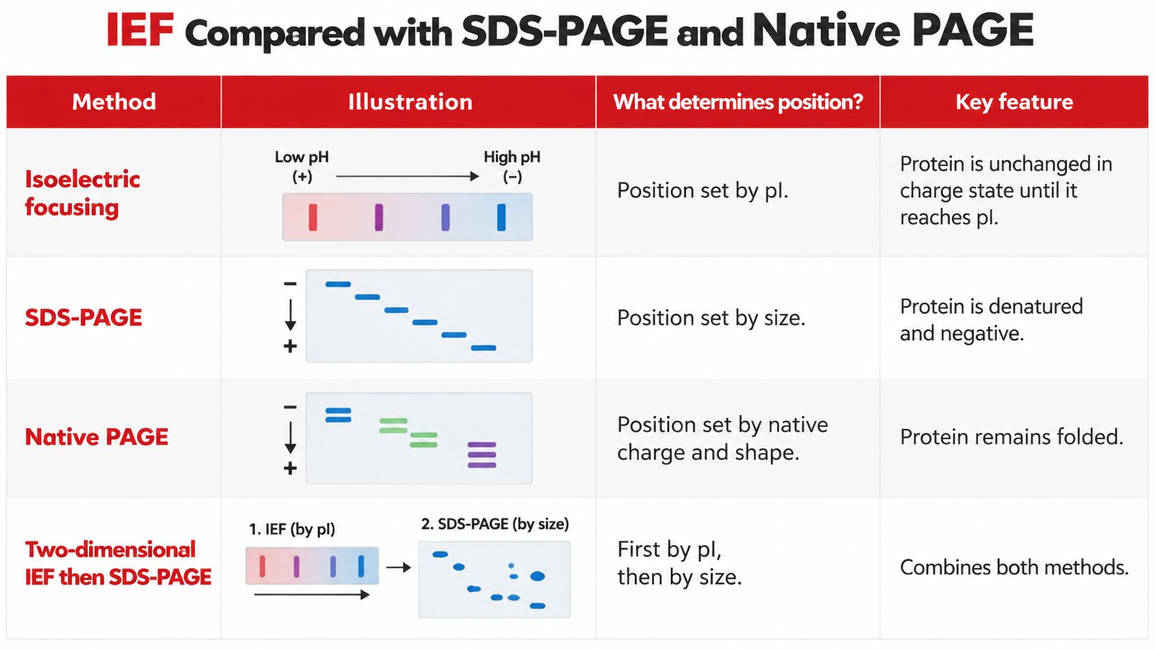
| Method | Position depends on | Protein state | Stem clue |
|---|---|---|---|
| Isoelectric focusing | pI, the pH of zero net charge | Ionizable groups remain; no SDS coat; may be native or denaturing | pH gradient, ampholytes, or pI values |
| SDS-PAGE | Molecular mass | Denatured and coated with negative SDS | SDS, subunits, or kilodalton ladder |
| Native PAGE | Both size and native charge, with shape also affecting mobility | Folded, no denaturant | Native conditions and no single-variable claim |
| IEF then SDS-PAGE | pI first, then mass | Native or urea IEF, then SDS in the second gel | Two dimensions or a 2D gel |
Sodium dodecyl sulfate–polyacrylamide gel electrophoresis (SDS-PAGE) and IEF answer different experimental questions. PAGE names the gel format; it doesn't, by itself, establish which molecular property determines separation.
Read the treatment before interpreting the bands
SDS denatures proteins and supplies a large negative charge approximately proportional to protein mass. This makes migration primarily a size-based comparison, with smaller proteins generally traveling farther through the gel.
Native PAGE doesn't impose that same denaturation and SDS coating. Protein size, native charge, and shape all contribute to mobility. A farther-traveling native protein can't automatically be called smaller without additional information.
IEF doesn't necessarily preserve folding. Denaturing IEF can use urea while still retaining the ionizable groups needed for pI-based focusing. The key distinction is the absence of the SDS charge treatment during the focusing separation.
The comparison graphic provides a visual reference, but the charge rule remains dynamic: a protein's charge changes as it encounters different pH regions during IEF.
Why combining methods reveals more
Proteins with matching pI values can occupy the same IEF band even when their masses differ. Following IEF with SDS-PAGE can separate those proteins along a new axis.
In 1975, P. H. O'Farrell reported this approach in High Resolution Two-Dimensional Electrophoresis of Proteins, published in the Journal of Biological Chemistry, volume 250, pages 4007–4021, using IEF first by pI and SDS-PAGE second primarily by mass. The experimental advantage is independent information about charge behavior and size.
A spot's location in such an experiment has meaning along both axes. Separation along the IEF axis suggests different pI values; separation along the SDS-PAGE axis suggests different apparent molecular masses.
The existing SDS-PAGE versus gel electrophoresis comparison covers size-based gels in more detail.
Decision line: A stem with a pH gradient or focusing positions calls for IEF logic. A stem with SDS and molecular weights calls primarily for mass logic; native conditions require attention to several properties.
Migration Traps and pI Shortcuts
Does movement toward an electrode mean that the protein's final band forms at that electrode? No. The BMJ account of isoelectric focusing supports the distinction between charge-driven migration and focusing where electrophoretic mobility reaches zero.
The same protein can start in different places and reach the same final band. The following study image accompanies an original scenario, not an official examination item.

Same protein, opposite initial directions
For a hypothetical pH 3 to 10 gradient, consider a protein with pI 7 loaded separately at different locations:
| Starting location | Initial interpretation | Final focused location |
|---|---|---|
| pH 4 | Net positive; moves toward the cathode | pH 7 |
| pH 9 | Net negative; moves toward the anode | pH 7 |
An original text diagram makes the opposing initial movements visible:
Positive anode                             Negative cathode
Acidic end                                      Basic end

pH 3      pH 4             pH 7       pH 9        pH 10
           positive  →   [band]   ←  negative
Suppose diffusion displaces a molecule from that band to pH 6.8. It becomes slightly positive and moves back toward the more basic region. At pH 7.2, it becomes slightly negative and moves back toward the more acidic region.
A protein doesn't need to cross its pI during ordinary directed migration. Crossing can occur through diffusion or displacement, after which the charge change provides the restoring movement.
What residue counting misses
A protein's pI isn't a simple average of its numbers of acidic and basic residues. Terminal groups, neighboring chemical groups, sequence context, and modifications affect how ionizable sites gain or lose protons.
That makes residue counting a rough qualitative clue rather than a complete calculation. Similar counts don't establish identical ionization behavior.
- For supplied pKa values: Identify the relevant charge states and the interval containing the net-zero form before applying a simplified pI calculation.
- For supplied pI values: Compare the stated local pH directly with pI instead of reconstructing the sequence.
- For mutations: Consider whether the substitution changes ionization, rather than assuming a small mass change means no band shift.
A useful mnemonic remains low pH, lots of protons, positive, cathode. It predicts the local charge and direction, not the distance traveled or the final endpoint at an electrode.
Worked IEF Passage and Review Checklist
An original protein-separation passage
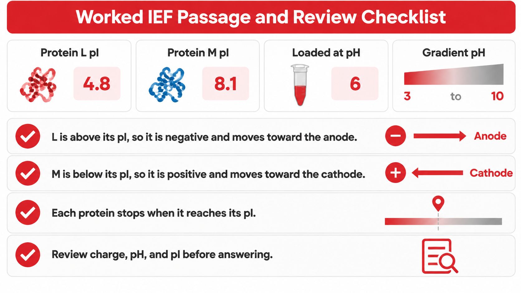
A hypothetical experiment uses a pH 3 to 10 gradient. Protein L has pI 4.8, Protein M has pI 8.1, and both are loaded at pH 6.
The starting pH lies above L's pI but below M's pI. The proteins therefore begin with opposite net charges even though they enter the same location.
| Protein | Comparison at loading | Initial charge and direction | Final position |
|---|---|---|---|
| L | pH 6 is above pI 4.8 | Negative; toward the anode | pH 4.8 |
| M | pH 6 is below pI 8.1 | Positive; toward the cathode | pH 8.1 |
Their masses aren't needed to assign these IEF positions. If the same proteins have equal masses and undergo standard SDS-PAGE, the pI difference that separated them in IEF won't itself separate them in that size-based experiment.
The worked-example graphic collects the starting conditions and reasoning.
An original multiple-choice question
A hypothetical protein with pI 6.0 is placed at pH 8.5 within an appropriate IEF gradient. Which choice correctly identifies its initial charge, initial direction, and final position?
- A. Positive; toward the cathode; stationary at pH 6.0.
- B. Negative; toward the anode; stationary at pH 6.0.
- C. Negative; toward the anode; stops at the anode.
- D. Negative; toward the anode; final position depends primarily on whether it's smaller than neighboring proteins.
B is correct. The initial pH exceeds the protein's pI, so the protein begins net negative. It moves toward the anode until it reaches the internal region matching its pI.
Choice A reverses the charge comparison. Choice C confuses initial electrode direction with final location, while choice D imports size-based reasoning from another separation method.
The peer-reviewed protein-separation article archived in PubMed Central supports the broader interpretation that charge differences can shift IEF bands even when size changes little. Conversely, equal pI values can produce coincident focusing positions despite different masses.
A reversed electrode is a fixable reading error, not evidence that biochemistry is out of reach. Recording the mistaken step makes the next review more useful than simply rereading the answer.
Additional MCAT biochemistry practice questions can help test whether the reasoning transfers beyond a familiar diagram.
A five-item passage-review checklist
- Identify the method. Look for a pH gradient, SDS treatment, or native conditions.
- Record each pI. Keep protein identities and charge properties separate.
- Compare starting pH with pI. Assign the initial net charge before predicting motion.
- Label the electrodes. Cathode is negative; anode is positive in this setup.
- Separate movement from destination. Write the initial direction and final focusing pH as different answers.
Plan the Next MCAT Biochemistry Session
A productive review session tests reasoning with changed conditions rather than repeating a memorized diagram. The MCAT study-plan guide can help place that review within a broader preparation schedule.
A self-check before seeking additional help
- Change the loading point: Rework the hypothetical L and M experiment from a new starting location within the gradient. Record each protein's charge, electrode direction, and final pH.
- Change the method: Explain aloud why matching masses would prevent the pI difference alone from separating those proteins on SDS-PAGE.
- Classify the error: Distinguish a charge mistake, an electrode-label mistake, and a method-selection mistake.
- Choose the next task: Use the electrophoresis comparison for a method gap or original passage questions for an interpretation gap.
For premeds also managing a thesis, Thesis Book Project's guidance to build a workable thesis roadmap addresses that separate writing workload. A thesis roadmap shouldn't substitute for passage practice or an MCAT-specific study plan.
Ace Med Boards offers private online MCAT tutoring covering content gaps, passage reasoning, timing, and retakes. Students share their test date, current practice results, available study time, and hardest preparation topics; current package information and terms appear on the MCAT pricing page.
Tutoring complements AAMC practice materials and independent study. It doesn't promise a particular score or admission outcome.
Sources
The scientific references linked within the sections above serve different purposes, so the most useful one to revisit depends on which step of the focusing mechanism is still unclear.
- Stanford's IEF overview: Describes the pH gradient, charge-dependent migration, and the focusing principle that carries each protein to the position where the local pH equals its pI.
- O'Farrell's Journal of Biological Chemistry paper: Documents the combination of pI-based focusing in one dimension and mass-based SDS-PAGE in the second.
- The BMJ account: Supports interpretation of migration and focusing behavior within the gradient.
- The Methods in Enzymology account: Provides context for ionization behavior and gradient design.
- The protein-separation article archived in PubMed Central: Supports interpreting charge-related differences between proteins that have focused at different positions in the gradient.
For a charge-sign or electrode-direction question, the Stanford overview is the fastest review. For proteins that share a mass but differ in pI, the O'Farrell paper and the protein-separation article show why pI and mass act as separate axes of separation.
Ace Med Boards offers private online MCAT tutoring that can connect protein chemistry with passage reasoning, including the distinction between initial migration and final focusing position. If guided passage review would help, a student can visit Ace Med Boards to schedule a free consultation and discuss preparation needs.