

You're reviewing a cardiology chapter, recognizing isolated facts, and then missing the question because the vignette asks for a physiological explanation rather than a label. Cardiology for medical students is best learned as a repeatable reasoning process: identify the presentation, localize the problem, connect it to hemodynamics or electrophysiology, choose the first diagnostic step, and triage urgency.
That approach applies across USMLE, COMLEX-USA, and Shelf-style questions. It also helps you understand why a murmur changes with preload, why an ECG localizes ischemia, and why a seemingly stable patient may need immediate escalation. This guide builds those connections from cardiac physiology to board-style clinical reasoning.
What Cardiology Really Tests on the Boards
A vignette about a 58-year-old man with crushing substernal pressure, diaphoresis, and left-arm numbness isn't testing whether you can recite every cause of chest pain. It compresses a clinical encounter into a few lines and asks you to integrate symptom recognition, coronary physiology, electrocardiography, biomarkers, and triage.
The most reliable way to approach cardiology questions is to use three skill axes:
- Pattern recognition: Identify familiar constellations such as ST-segment elevation myocardial infarction, atrial fibrillation, or a classic valve murmur.
- Mechanism reasoning: Explain why the finding occurs. An S3 suggests rapid ventricular filling into a volume-overloaded ventricle, while exertional syncope in aortic stenosis reflects a fixed obstruction that limits the rise in cardiac output.
- Management triage: Decide whether the patient needs emergent reperfusion, urgent stabilization, procedural evaluation, or outpatient testing.
Memorization helps you recognize the topic, but it won't reliably identify the best answer. The same underlying problems, reduced coronary blood flow, impaired ventricular filling, abnormal conduction, or obstructed forward flow, appear in different clinical settings.
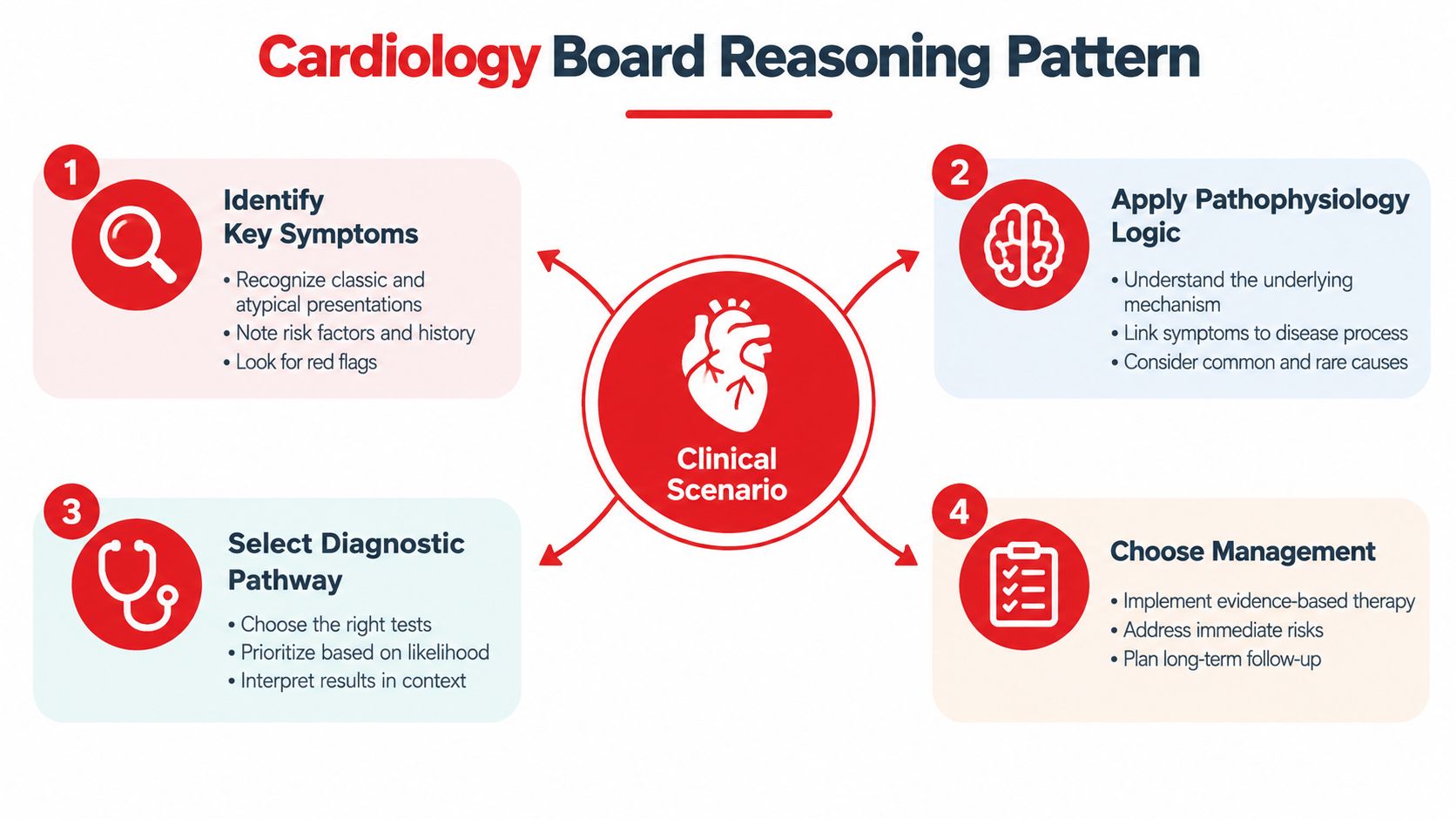
A four-step reading habit
When you open a cardiac vignette, ask:
- What is the dominant presentation? Chest pain, dyspnea, syncope, palpitations, or edema?
- Where is the abnormality? Coronary territory, valve, myocardium, conduction system, pericardium, or great vessel?
- What mechanism explains the findings together? Use pressure, volume, oxygen supply, electrical conduction, or obstruction.
- What action matches the risk? Stable patients permit confirmation. Unstable patients require simultaneous diagnosis and intervention.
The USMLE content outline can help you organize these topics by the kind of knowledge boards assess, rather than by an unstructured disease list. For real clinical practice, diagnosis and treatment require complete history, examination, testing, and supervision. This article is for examination education, not individualized medical advice.
Core Anatomy and Physiology You Must Know
Cardiology becomes easier when anatomy explains the ECG and physiology explains the symptom. Start with the coronary territories. In a right-dominant circulation, the right coronary artery commonly supplies the inferior wall and the sinoatrial node, the left anterior descending artery supplies the anterior wall and apex through its diagonal branches, and the left circumflex artery supplies much of the lateral wall.
| Coronary Artery | Wall Supplied | ECG Leads | Classic Association |
|---|---|---|---|
| Left anterior descending artery | Anterior wall and apex | V1 through V4 | Anterior ischemia or infarction |
| Right coronary artery | Inferior wall and often the sinoatrial node | II, III, and aVF | Inferior infarction, possible bradyarrhythmia |
| Left circumflex artery | Lateral wall | I, aVL, V5, and V6 | Lateral ischemia or infarction |
The four valves create pressure gradients that determine when they open. The atrioventricular valves open when atrial pressure exceeds ventricular pressure, while the semilunar valves open when ventricular pressure exceeds arterial pressure. A stenotic valve preserves forward flow only by requiring a greater upstream pressure, whereas regurgitation sends blood backward and increases volume load.
From ventricular function to bedside findings
The Frank-Starling relationship describes how increased ventricular filling can increase contractile force within physiological limits. In systolic dysfunction, the ventricle can't generate adequate pressure or ejection. In diastolic dysfunction, the ventricle may eject a relatively preserved fraction but fills poorly because it is stiff.
A failing left ventricle raises left ventricular end-diastolic pressure. That pressure backs up into the left atrium and pulmonary circulation, producing pulmonary crackles and dyspnea. If the process also increases systemic venous pressure through broader heart-failure physiology, the student may see jugular venous distention and peripheral edema. An S3 gallop reflects rapid filling into a volume-loaded ventricle, so the sound is a hemodynamic clue, not an isolated memorization item.
Autonomic physiology adds another layer. Vagal maneuvers increase parasympathetic influence through the atrioventricular node and can interrupt some reentrant supraventricular tachycardias. Beta-adrenergic blockade reduces sympathetic stimulation and myocardial oxygen demand, which explains its role in selected post-infarction contexts, although real-world treatment depends on the patient's stability and contraindications. For a more focused review, use this cardiovascular physiology review.
Worked physiology example
An older adult with exertional syncope and a harsh systolic ejection murmur may have a narrowed aortic valve. During exercise, the body needs cardiac output to rise, but the fixed obstruction limits forward flow. The ventricle generates higher pressure to eject blood, myocardial oxygen demand increases, and cerebral perfusion can fall. The board-style chain is fixed outflow obstruction, inability to augment output, exertional syncope, and demand ischemia.
High-Yield Cardiac Pathologies Compared
The fastest way to distinguish cardiac diseases is to compare presentation, first test, and urgency. Don't let a shared symptom such as dyspnea erase the difference between pulmonary congestion from heart failure, ischemia, arrhythmia, or pulmonary embolism.
| Pathology | Classic Presentation | First Test | Initial Triage |
|---|---|---|---|
| Stable angina | Predictable exertional pressure relieved by rest | ECG and risk-based ischemic evaluation | Outpatient evaluation if stable |
| NSTEMI | Ischemic symptoms with myocardial injury but no diagnostic ST elevation | Serial troponin and ECG | Risk stratification and monitored care |
| STEMI | Acute ischemic symptoms with diagnostic ST elevation | Immediate ECG | Emergent reperfusion pathway |
| Heart failure with reduced ejection fraction | Dyspnea, orthopnea, edema, and systolic dysfunction | Natriuretic peptide and echocardiography | Stabilize if decompensated, then identify cause |
| Heart failure with preserved ejection fraction | Congestion with impaired relaxation and a relatively preserved ejection fraction | Natriuretic peptide and echocardiography | Assess congestion and precipitating factors |
| Aortic stenosis | Exertional syncope, angina, or dyspnea with a systolic ejection murmur | Echocardiography | Urgent specialty assessment when symptomatic |
| Aortic regurgitation | Wide pulse pressure and diastolic decrescendo murmur | Echocardiography | Assess severity and ventricular response |
| Mitral stenosis | Dyspnea, atrial arrhythmia, and an opening snap | Echocardiography | Evaluate obstruction and rhythm |
| Mitral regurgitation | Holosystolic murmur radiating toward the axilla | Echocardiography | Escalate urgently if acute pulmonary edema develops |
| Atrial fibrillation | Irregularly irregular rhythm and absent organized P waves | Rhythm strip or ECG | Rate control if stable, urgent cardioversion if unstable |
| Supraventricular tachycardia | Regular narrow-complex tachycardia with sudden onset | ECG | Vagal maneuvers or adenosine in an appropriate stable rhythm |
| Ventricular tachycardia | Wide-complex tachycardia, often in structural heart disease | ECG | Treat as dangerous until proven otherwise |
| Torsades de pointes | Polymorphic ventricular tachycardia with prolonged QT | ECG | Magnesium and defibrillation when unstable |
| Congenital shunt or coarctation | Murmur, differential pulses, cyanosis, or a fixed splitting pattern | Echocardiography and targeted imaging | Match urgency to hemodynamic effect |
Contrasts that change the answer
STEMI and NSTEMI aren't interchangeable. ST elevation with an appropriate ischemic presentation triggers an emergent reperfusion pathway. NSTEMI requires biomarker interpretation, monitoring, and risk stratification rather than automatic equivalence with STEMI.
Symptomatic aortic stenosis is a warning that fixed obstruction has become clinically consequential. Acute mitral regurgitation can instead present with abrupt pulmonary edema and requires urgent evaluation for a mechanical cause. In rhythm questions, stable atrial fibrillation with rapid ventricular response is approached differently from unstable tachycardia, and torsades requires magnesium and electrical defibrillation when indicated, not routine treatment as though it were ordinary atrial fibrillation. Review the distinctions in this acute coronary syndrome resource.
Reading ECGs With a Board-Style Framework
An ECG becomes less intimidating when you read it in the same order every time. Use rate, rhythm, axis, intervals, morphology, and ST/T changes. This prevents one striking feature from causing premature closure.
The systematic sweep
Begin with the ventricular rate, then decide whether the rhythm is regular. Look for P waves and ask whether each P wave is followed by a QRS complex. Next, assess the axis, followed by the PR interval, QRS width, and QT interval. Only then interpret QRS morphology, R-wave progression, ST segments, and T waves.
Use a labeled composite strip for practice rather than treating every ECG as a flashcard. For example, label the strip rate, rhythm, axis, PR, QRS, QT, ST segment, and T wave. Hide the diagnosis and write one observation beside each label. This separates what you see from what you infer.
Practical rule: A diagnosis should explain the rhythm, the interval abnormality, and the clinical context together.
| Pattern | Key ECG Finding | Board Pearl |
|---|---|---|
| Anterior STEMI | ST elevation across anterior precordial leads | Think anterior wall injury and LAD territory |
| Inferior STEMI | ST elevation in II, III, and aVF | Check for right-ventricular involvement when the presentation supports it |
| Wellens pattern | Deeply inverted or biphasic T waves in the anterior leads during a pain-free period | Signals critical proximal LAD disease and is not a benign outpatient ECG finding |
| Brugada pattern | Characteristic ST-segment abnormality in the right precordial leads | Connect the pattern with ventricular arrhythmia risk and sodium-channel physiology |
| Hyperkalemia | Progressive peaked T waves, conduction slowing, and possible QRS widening | Interpret the ECG as an electrical emergency when the clinical context fits |
| Acute pericarditis | Diffuse ST elevation with reciprocal changes typically limited to aVR | Distinguish diffuse inflammation from a regional coronary territory |
| QT prolongation | Extended QT interval | Ask about medications, electrolyte disturbance, and torsades risk |
The point isn't to memorize shapes without context. Inferior ST elevation reflects regional myocardial injury, diffuse ST elevation suggests a broader inflammatory process, and QT prolongation lengthens repolarization and permits polymorphic ventricular arrhythmia. Practice with these ECG reading steps until the sequence feels automatic.
Diagnostic Workup and Management Algorithms
Board vignettes often reveal the correct pathway through the order of information. A patient with chest pain or dyspnea should first be classified as stable or unstable, then evaluated through an ischemic or nonischemic branch.
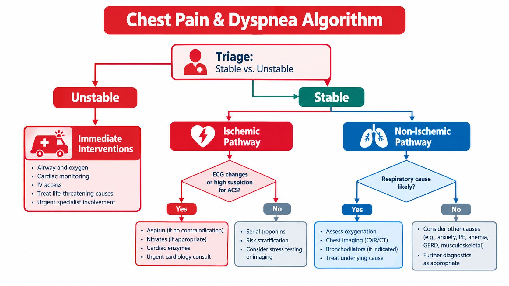
Start with triage
For an unstable patient, prioritize airway, breathing, circulation, monitoring, intravenous access, and immediate senior or procedural support while collecting focused data. Hypotension, altered mental status, severe hypoxemia, malignant arrhythmia, or signs of cardiogenic shock should move the question away from leisurely outpatient testing.
For a stable patient, obtain a focused history and examination, ECG, cardiac biomarkers when ischemia is plausible, and targeted imaging. A chest radiograph can support pulmonary or structural reasoning, while bedside echocardiography can reveal ventricular dysfunction, pericardial fluid, right-heart strain, or gross valve pathology.
Use the right branch
For suspected acute coronary syndrome, interpret the ECG promptly and use serial troponin testing rather than relying on a single result. STEMI generally moves toward urgent reperfusion and catheter-based evaluation when available. Fibrinolysis is a time-sensitive alternative only in the appropriate setting when immediate percutaneous intervention isn't available and contraindications have been assessed.
For acute decompensated heart failure, evaluate congestion, oxygenation, perfusion, precipitating ischemia, rhythm, infection, and medication or dietary factors. Natriuretic peptides are useful rule-out tests in symptomatic patients. The ESC thresholds cited in guideline summaries are BNP 35 pg/mL and NT-proBNP 125 pg/mL, with low values making heart failure less likely, while high values require context because age, renal dysfunction, atrial fibrillation, obesity, and acute coronary syndromes can affect interpretation. These thresholds are summarized in the European Journal of Heart Failure practical guidance.
If pulmonary embolism remains plausible, use pretest probability to decide whether D-dimer is appropriate. CT pulmonary angiography is used when imaging is indicated and feasible, while lower-extremity Doppler can support venous thromboembolism evaluation in selected patients.
A cardiovascular risk panel, such as this private blood test from Repose Healthcare, belongs to preventive-risk discussions, not to the emergency evaluation of crushing chest pain, dyspnea, or suspected infarction.
Walking Through a Composite Cardiology Vignette
The following is a labeled composite educational example, not a real Ace Med Boards student case and not a recalled examination question. A 58-year-old man arrives with crushing substernal pain, diaphoresis, and dyspnea.
His blood pressure is low, his pulse is slow, and his lungs are relatively clear. The first move is not to jump directly to a named infarct. First, identify instability. Then obtain an ECG, establish monitoring and access, assess perfusion, and consider right-sided leads because the examination suggests an inferior process with possible right-ventricular involvement.
Localize before treating
The ECG shows ST elevation in leads II, III, and aVF, with greater elevation in III than II. That localizes injury to the inferior wall and points toward the right coronary territory. A right-sided lead, V4R, also shows ST elevation, supporting right-ventricular extension.
The physiology now explains the apparently unusual combination of hypotension, bradycardia, and clear lungs. Right-ventricular dysfunction reduces delivery of blood to the left ventricle, lowering left-sided preload and cardiac output. Inferior infarction can also affect nodal blood supply, which helps explain the slow pulse.
Exam reasoning: Don't treat the ECG in isolation. Let the blood pressure, lung examination, rhythm, and lead distribution reinforce one mechanism.
A classic distractor is a nitrate in a preload-dependent right-ventricular infarction. Reducing venous return can worsen hypotension, so the student must recognize the hemodynamic context before selecting an intervention. Another distractor is fibrinolysis for a late-presenting STEMI when the stem instead supports transfer for urgent procedural evaluation, or when contraindications make it inappropriate. The correct answer depends on timing, resources, contraindications, and stability, not on a memorized medication list.
The next danger is a mechanical complication. New hypotension with a new harsh murmur, abrupt pulmonary edema, a new ventricular septal defect, or acute severe mitral regurgitation should shift the answer toward urgent echocardiography and specialist or surgical escalation. The reasoning chain is presentation, stability, ECG localization, right-sided physiology, immediate reperfusion pathway, and complication surveillance.
Here's a short visual review of the clinical reasoning sequence before you replay the vignette:
For any new cardiology vignette, ask the same five questions: What is the presentation? Where is the lesion? What physiology links the findings? What test confirms the pathway? Which red flag changes the disposition?
Board Study Plan and Practice Question Strategy
A focused cardiology review works better when each week has a defined job. Use a four-week sprint built around your required core text, a single high-quality question bank, and repeated ECG and hemodynamic retrieval.
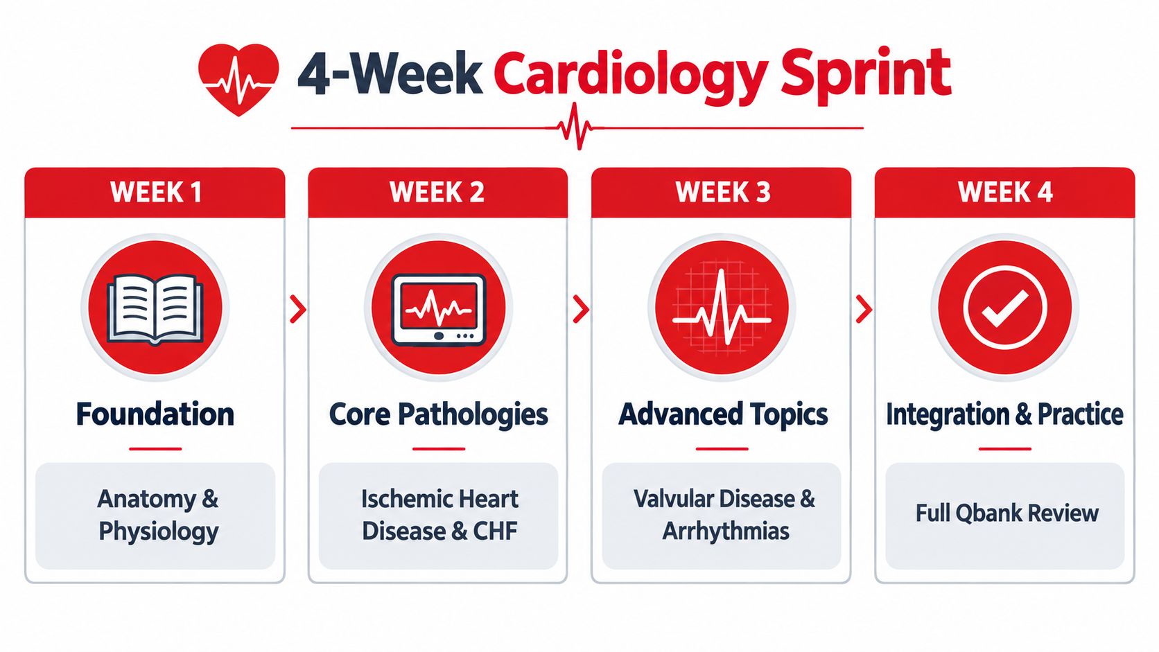
Four weeks with a purpose
| Week | Main Focus | Output to Produce |
|---|---|---|
| Week 1 | Cardiac anatomy and physiology | Pressure-flow map and coronary lead table |
| Week 2 | Ischemic heart disease and heart failure | ACS and congestion decision trees |
| Week 3 | Valvular disease and arrhythmias | Murmur comparison sheet and ECG pattern log |
| Week 4 | Congenital disease, pericardial disease, and integration | Mixed question review and error audit |
A daily routine can be simple: read a manageable assigned portion, complete targeted questions, and review every item actively. The exact page and question count should fit your schedule and current baseline. What matters is that review takes longer than passive answer checking.
For each missed question, write one sentence containing the mechanism, the discriminating clue, and the next step. Tag the miss as knowledge gap, reasoning error, misread stem, or premature closure. Then link it to a related concept. A question about right-ventricular infarction may connect to preload, venous return, inferior ECG localization, and nitrate physiology.
Mnemonics need explanation
Use mnemonics only after you understand the mechanism. SALAMI can help organize ischemic ECG evolution, MADNESS can prompt a broad syncope differential, and SATCHEL can support chest-pain localization. Don't let the mnemonic replace the actual question, which is usually asking why the patient has the finding or what must happen next.
Spaced retrieval is more useful when you revisit the same concept after an interval and attempt to reconstruct it before looking at notes. This spaced repetition guide can fit into a schedule that includes physiology, question review, and ECG practice.
On Shelf or board-style testing days, pace by blocks rather than by anxiety. Flag uncertain items, record the reason for the uncertainty, and return only after completing the remaining questions. If you must guess, translate the guess into a mechanism-based choice: identify the unstable feature, the lesion's location, and the intervention that addresses the immediate threat.
Educational support should remain individualized to the learner's actual exam, baseline, and available time. Ace Med Boards offers diagnostic tutoring and personalized study-plan support for board preparation, so students who want structured help can visit Ace Med Boards to discuss whether that format fits their cardiology review.
Key Takeaways
- Think in mechanisms: Connect symptoms to pressure, volume, oxygen supply, obstruction, or conduction.
- Read ECGs systematically: Rate, rhythm, axis, intervals, morphology, and ST/T changes prevent premature closure.
- Triage before treatment: Stability determines whether the vignette calls for confirmation, monitored care, urgent intervention, or escalation.
- Review errors by type: Separate missing knowledge from faulty reasoning so your next study block addresses the actual problem.
Educational note
This article is intended for examination education for medical students, residents, applicants, and graduates preparing for USMLE, COMLEX-USA, or relevant Shelf examinations. It isn't a diagnosis or a substitute for supervised clinical training, institutional protocols, or patient-specific medical advice.